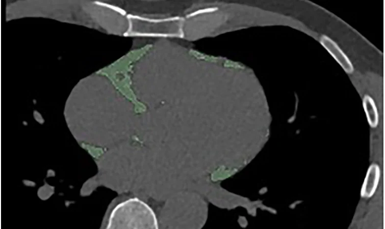

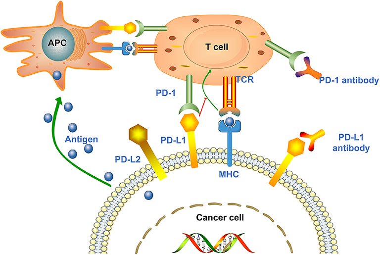

Welcome, fellow creatures of the radiology night. Tonight’s tale is a small one, but a tantalizing one: could a photon-counting CT (PCCT) scan hint at a lung tumor’s PD-L1 status without a single biopsy needle involved? Let us count the evidence carefully — because a weak correlation dressed up as a diagnosis is exactly the sort of number that keeps me up at night.
Key takeaways
- A single-center study of 63 non-small cell lung cancer (NSCLC) patients found two PCCT spectral parameters weakly but significantly correlated with PD-L1 expression [1][2].
- A combined clinical-plus-PCCT model reached an AUC of 0.889 for detecting high PD-L1 expression in this cohort — but with no external validation [1][2].
- This is hypothesis-generating, single-center, retrospective work; every patient’s PD-L1 status was still determined by biopsy, which remains the standard [1][2].
- The findings add to a small, growing literature linking spectral or dual-energy CT features to PD-L1 status, now extended to true photon-counting hardware [3][5][6][8].
Why This Study Matters
PD-L1 expression, measured on biopsy tissue via immunohistochemistry, helps guide whether NSCLC patients might benefit from immune checkpoint inhibitor therapy. Because biopsy is invasive and can be limited by tumor heterogeneity or sampling, researchers have long looked for imaging-based ways to estimate PD-L1 status non-invasively. This new study, published July 1, 2026, in BMC Medical Imaging, tests whether photon-counting CT can add a quantitative “spectral fingerprint” to that search [1][2].
Study Design: What They Actually Did
Researchers at Shandong Cancer Hospital and Institute in China retrospectively analyzed pretherapeutic PCCT scans from 63 NSCLC patients, obtained before any treatment. Biopsy-based immunohistochemistry classified PD-L1 expression into three tiers — negative, intermediate, and high — using the tumor proportion score (TPS) [1][2].
From the PCCT scans, the team measured a set of spectral parameters in both arterial and venous phases: CT attenuation at 40, 70, and 100 keV monoenergetic reconstructions, lesion iodine concentration, normalized iodine concentration, and the slope of the spectral Hounsfield-unit curve (λHU) [1][2]. They then built three logistic regression models — clinical variables alone, PCCT parameters alone, and a combined model — and compared them by ROC analysis [1][2].
What They Found
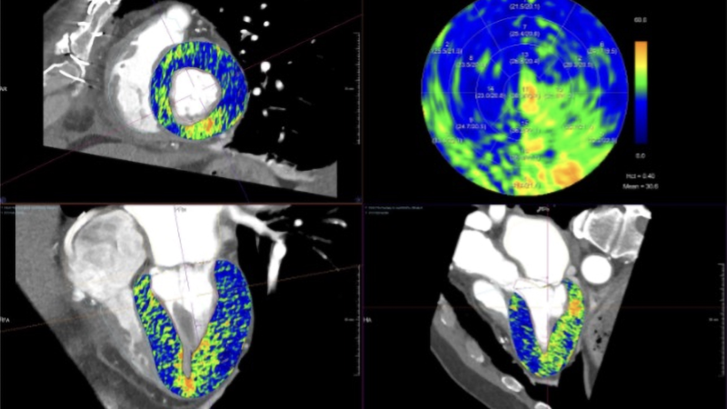
Of all the parameters tested, only two reached statistical significance: arterial-phase 40 keV attenuation (A-40 keV) and the arterial spectral-curve slope (A-λHU). Both showed weak negative correlations with PD-L1 expression — A-λHU: rs = −0.270, P = 0.032; A-40 keV: rs = −0.279, P = 0.027 [1][2]. In multivariate regression, age (OR = 1.014) independently predicted intermediate PD-L1 expression, and A-λHU (OR = 0.031) independently predicted high PD-L1 expression [1][2].
For distinguishing high PD-L1 expression, ROC analysis gave an AUC of 0.762 (95% CI 0.637–0.860) for the clinical-only model, 0.846 (95% CI 0.733–0.925) for the PCCT-only model, and 0.889 (95% CI 0.785–0.954) for the combined model (all P < 0.001) [1][2]. The combined model performed best in this cohort.
I have spent four centuries counting grains of rice and cobblestones, and an r-value of −0.27 is, to me, a very small pile indeed. Statistically real, yes. Clinically decisive, not yet.
Reading the Result Honestly
This is a single-center, retrospective study of 63 patients — near the bottom of the evidence ladder for clinical relevance, even though it is peer-reviewed rather than a preprint or conference abstract [1][2]. The correlations reported are statistically significant but weak by conventional standards, and only two of many tested spectral parameters reached significance [1][2]. The AUC figures come from a single internal cohort with no held-out test set or external validation, and the confidence intervals are wide and overlapping [1][2].
This work sits within a small but growing family of studies using dual-energy or dual-layer spectral CT to probe PD-L1 status non-invasively [3][5][6][8]. What is genuinely new here is the use of true photon-counting detector hardware rather than dual-layer or dual-source dual-energy CT — extending an existing line of inquiry to a newer scanner class, not delivering a qualitative leap [1][2].
Limitations Worth Naming
- Small, single-center sample (N = 63) limits statistical power and generalizability [1][2].
- Retrospective design with waived informed consent; selection bias in which patients received both pretreatment PCCT and biopsy cannot be excluded [1][2].
- No external validation cohort or prospective test set is reported; the AUCs likely reflect in-sample performance [1][2].
- The manuscript is labeled by the publisher as an unedited pre-final version, so some methodological detail — exact scanner model, radiation dose, histology and stage breakdown, PD-L1 antibody clone — could not be confirmed from the accessible text [1][2].
- One co-author is affiliated with Siemens Healthineers; the study declares no competing interests, but the affiliation is worth noting given the vendor context for PCCT availability in China [1][2][19].
- PD-L1 status still required tissue biopsy in every patient studied — this describes a statistical association with imaging, not a replacement for biopsy [1][2].
What Would Change This Assessment
A larger, prospective, multicenter cohort with predefined imaging protocols would be the obvious next step, along with testing whether the model generalizes across NSCLC histologic subtypes and PCCT scanner models. A head-to-head comparison with existing dual-energy or radiomics approaches, in the same patients, would also help place this work in context. Eventually, a formal decision-analytic study could ask whether an imaging score might usefully triage which patients need biopsy re-testing — a modest, testable question, and a fair one.
What This Means for Practice Today
For imaging professionals, this study is best read as an early signal, not a new tool. The authors themselves suggest their combined model “may serve as a non-invasive tool for individualized treatment” [1][2] but that is their own interpretation of preliminary, single-center data, not an established clinical capability. Biopsy remains the standard for determining PD-L1 status and immunotherapy eligibility, and nothing in this study changes that.
FAQ
Can photon-counting CT replace biopsy for PD-L1 testing?
No. This study found a statistical association between certain PCCT spectral parameters and PD-L1 expression, but every patient’s PD-L1 status was still determined by tissue biopsy, and the researchers describe their model as a potential complement, not a replacement [1][2].
How strong is the correlation between PCCT parameters and PD-L1 expression?
Weak by conventional statistical standards, though statistically significant, with correlation coefficients (rs) of −0.27 to −0.28 [1][2].
Has this finding been confirmed at other centers?
Not yet. This is a single-center, retrospective study with no external or prospective validation reported [1][2].
Does this study use the same technology as earlier spectral-CT PD-L1 research?
It extends earlier work using dual-energy or dual-layer spectral CT [3][5][6][8] to true photon-counting detector CT, a newer scanner class, not a different underlying idea.
Could PCCT eventually predict immunotherapy response?
That is not what this study tested. It looked only at correlation with PD-L1 expression as measured by biopsy, not treatment outcomes.
References
1. Ren J, Fu Z, Li L, Huang Y, Yin Y. Quantitative spectral parameters of photon-counting detector CT for noninvasive prediction of PD-L1 expression in non-small cell lung cancer. BMC Med Imaging. 2026 Jul 1.
2. Same article, publisher page (BMC Medical Imaging / SpringerLink) — abstract, funding, and ethics/competing-interests statements.
3. Lou H, Cui S, Dong Y, et al. Identification of PD-L1 Expression in Resectable NSCLC using Interpretable Machine Learning Model Based on Spectral CT. Curr Med Imaging. 2025 Oct 9.
5. Predictive value of spectral dual-detector computed tomography for PD-L1 expression in stage I lung adenocarcinoma: development and validation of a novel nomogram.
6. Is there any correlation between spectral CT imaging parameters and PD-L1 expression of lung adenocarcinoma?
8. Multiparameter spectral CT-based radiomics in predicting the expression of programmed death ligand 1 in non-small-cell lung cancer.
19. The dawn of photon-counting CT and its clinical revolution in China (trade press; context on PCCT market availability, not a scientific source for this study’s findings).

Until the multicenter trial arrives, dear reader, I shall keep counting. Stay curious, stay skeptical, and I’ll see you in the reading room.
Want more findings counted honestly, hedges intact? Subscribe to The Photon Count newsletter.
Educational content, not medical advice. Count Photon explains imaging technology and published research for educational purposes. Nothing here is medical advice, and it should never replace a conversation with a qualified healthcare professional about your own care.