
What has changed since our 2025 report
When we first covered this field, clinical photon-counting CT (PCCT) was essentially a one-scanner story, and most of the evidence came from single-centre feasibility work. Eighteen months on, enough has moved that a full rewrite made more sense than a patch.
Three developments stand out:
- PCCT is no longer a single-vendor technology. For its first few years, clinical cardiovascular PCCT meant one platform, the dual-source Siemens NAEOTOM Alpha, cleared by the FDA in 2021. That changed in 2026. GE HealthCare won FDA 510(k) clearance for its Photonova Spectra system on 23 March 2026, built on a new “Deep Silicon” detector with 8-bin energy resolution, and dosed its first patient in an international multicentre trial a month later, on 23 April. A portable photon-counting extremity scanner from MARS Bioimaging was also cleared in March 2026.
- The evidence grew up. We now have systematic reviews and pooled analyses of coronary PCCT, so for the first time it’s possible to talk about summary estimates rather than one institution’s numbers.
- The clinical question widened. Work in 2025 and 2026 pushed PCCT past stenosis, stent and plaque assessment and into the myocardium itself, using late enhancement, extracellular volume and perfusion to get tissue information that used to require MRI.
The sections below keep the cardiovascular focus of the original but revisited each claim to the latest 2026 evidence. New for this edition, we will also close with a section of what the technology still hasn’t shown.
Where the technology stands in 2026
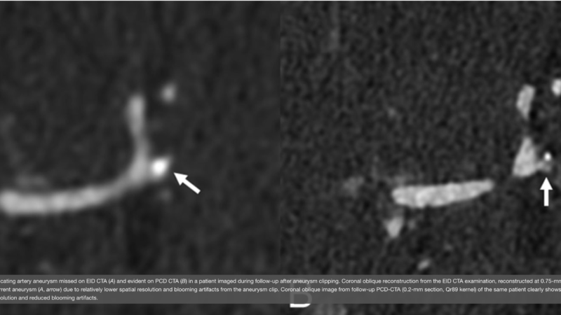
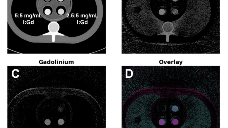
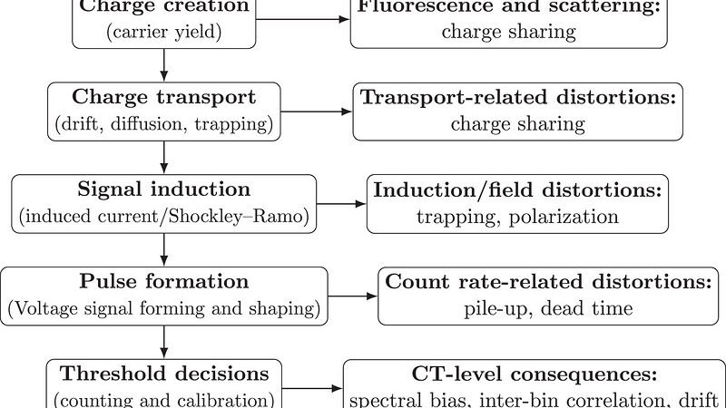
PCCT detectors turn individual X-ray photons directly into electrical signals whose size tracks photon energy. Conventional energy-integrating detectors (EID) instead pool light from a scintillator. Every recent review lists the same cardiovascular payoffs from that difference: ultra-high spatial resolution, spectral (multi-energy) data on every scan, less electronic noise and blooming, a stronger iodine signal, and better dose efficiency.
What’s actually new is access. A second clinical cardiovascular platform, GE’s silicon-based system, now sits alongside the established Siemens dual-source design. Almost all of the findings below were generated on the NAEOTOM Alpha, so the obvious next test is whether they hold up on a different detector material and a different vendor. For my money, that cross-platform question is the one to watch over the next year or two.
Cardiovascular imaging: the evidence
1. Coronary stents and in-stent restenosis
The in-stent lumen has always been one of CCTA’s blind spots, because the metal struts bloom and swamp the small lumen underneath. This is where UHR PCCT earns its keep.
Hagar et al. (2024) prospectively assessed 44 stents in 18 patients (mean age 83) in UHR mode at 120 x 0.2 mm collimation. Against invasive coronary angiography (ICA) they reported 100% sensitivity and 92.3% specificity, with only 3 of 44 stents non-diagnostic. (Eur Radiol; DOI: 10.1007/s00330-023-10516-3)
Qin et al. (2024) compared standard and ultra-high-resolution protocols across 131 stents. UHR accuracy came in at 88.0% versus 78.3% for standard resolution, and the authors settled on sharp vascular kernels (Bv72 or Bv76) at 0.2 mm as the sweet spot. (Eur Radiol; DOI: 10.1007/s00330-024-10760-1)
Shin, Sakai et al. (2025) is a useful newer addition, and a larger one. Across 283 stented lesions in 171 patients, measured against quantitative coronary angiography, PCD-CT reached 80.0% sensitivity, 90.4% specificity, 96.4% NPV and 88.9% overall accuracy for obstructive in-stent restenosis, with only 1.1% of lesions indeterminate. (EuroIntervention 2025;21(19):e1137-e1146; DOI: 10.4244/EIJ-D-25-00257)
Boccalini et al. (2022) is still the reference first-in-human spectral PCCT stent study, on a separate Philips research prototype (SPCCT), showing less blooming and clearer lumen than dual-layer CT. (Invest Radiol; DOI: 10.1097/RLI.0000000000000835)
Worth pairing those best-case numbers with the pooled picture, though. A 2026 systematic review found that once you combine studies, UHR PCD-CT performance for in-stent stenosis was only moderate (sensitivity around 62.6%, specificity around 62.4%), which reflects how much stent type, size and protocol vary between series. PCCT plainly improves stent imaging. The “100% sensitivity” headline just belongs to favourable, well-chosen cohorts rather than to the average patient.
2. Plaque characterisation
By combining UHR with spectral data, PCCT separates calcified, fibrous and lipid-rich components more cleanly, and it cuts the blooming that inflates how much calcified plaque there looks to be.
Mergen et al. (2022), the first in-human quantitative UHR plaque study, ran 20 patients and found 6.7% lipid-rich components versus 0.5% with standard imaging, while the apparent calcified-plaque fraction dropped from 85.1% to 75.2% once blooming was reduced. (Front Cardiovasc Med; DOI: 10.3389/fcvm.2022.981012)
Dahal et al. (2024) checked PCCT against histology and could tell haemorrhage from fibrous cap (p=0.017), lipid (p=0.003) and necrosis (p=0.004), and thrombus from fibrosis (p=0.048). (Eur Radiol; DOI: 10.1007/s00330-022-09155-x)
Vattay et al. (2024) showed that the virtual monoenergetic level you choose (40 to 70 keV) really does change measured plaque attenuation and volume, and that 70 keV gave the smallest relative difference for calcified-plaque quantification. (Eur Radiol; DOI: 10.1007/s00330-023-09876-7)
Si-Mohamed et al. (2022) reported a 2.3-fold higher detectability index for the coronary lumen and 2.9-fold for non-calcified plaque against conventional CT. (Radiology; DOI: 10.1148/radiol.211780)
Vecsey-Nagy et al. (2025) added an intra-individual comparison of UHR PCD-CT against EID-CT for plaque quantification, confirming that PCCT reports systematically lower calcified-plaque volumes. (Radiology 2025;314(3):e241479; DOI: 10.1148/radiol.241479)
All of this now lives inside a broader policy frame. The 2025 ACC Scientific Statement on quantitative coronary plaque analysis (Chandrashekhar, Blankstein, Shaw et al., JACC: Cardiovascular Imaging, December 2025) sets out how quantitative plaque metrics should be used in practice, and that’s the yardstick PCCT plaque data will be measured against.
3. Ultra-high-resolution and calcified coronary disease
If there’s one reproducible cardiovascular win for PCCT, it’s cutting the false overestimation of stenosis severity that calcium blooming causes.
Emrich et al. (2024) found UHR PCCT tightened stenosis quantification (mean bias 2.3% versus 10.1% for standard resolution, P<0.001) and moved 54% of patients to a lower CAD-RADS category, with median calcified-plaque stenosis falling from 41.5% to 26.7% (P<0.001). (Radiology; DOI: 10.1148/radiol.231956)
Hagar et al. (2023) looked at 68 high-risk TAVR candidates with severe aortic stenosis and reached 96% sensitivity, 84% specificity and 88% accuracy for 50% or greater CAD (AUC 0.93), heavy calcification and all. (Radiology; DOI: 10.1148/radiol.223305)
Geering et al. (2023) documented the resolution that makes this possible: 0.11 x 0.11 mm in-plane and 0.16 mm through-plane. (J Cardiovasc Comput Tomogr; DOI: 10.1016/j.jcct.2023.02.009)
Kotronias et al. (2025) put the calcified-CAD claim through its stiffest test yet, benchmarking PCCT angiography against invasive assessment specifically in severely calcified coronaries. (JACC Cardiovasc Imaging 2025;18:572-585)
Alongside UHR, virtual non-calcium (VNCa) reconstructions and virtual calcium scoring from a single CCTA are now workable on PCCT, which gives another way to see past calcium without adding a non-contrast scan.
4. Myocardial tissue characterisation
This is probably the biggest change since our last write-up. Spectral PCCT now does myocardial characterisation that used to be MRI’s territory.
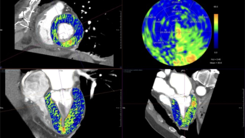
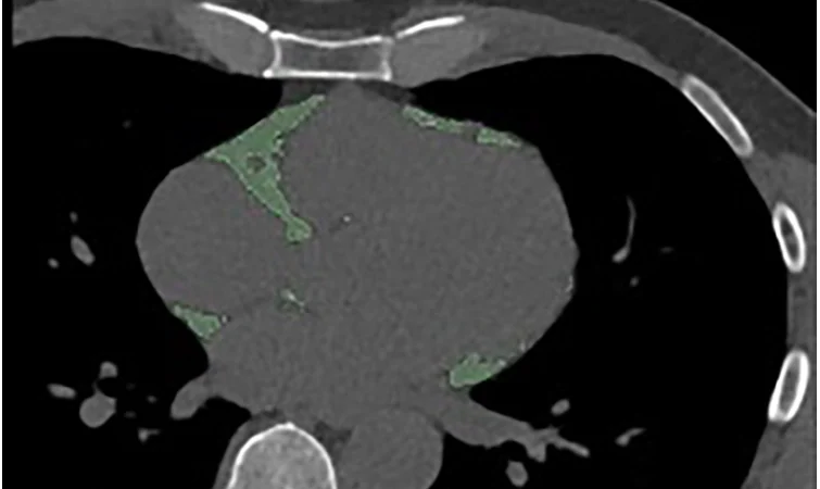
On late iodine enhancement, PCCT iodine maps pick up myocardial scar with good agreement against LGE MRI (Tremamunno et al., Eur Radiol 2025;35:7074-7083; Gnasso et al.). In spontaneous coronary artery dissection, late-enhancement PCD-CT agreed strongly with cardiac MRI, with kappa reaching 0.944 (Klambauer et al., Invest Radiol 2025).
On extracellular volume, spectral late-enhancement scans give ECV values that track MRI closely (reported r of roughly 0.82 to 0.91), which opens the door to diffuse fibrosis and infiltrative disease such as cardiac amyloidosis.
On arrhythmia, late-enhancement PCD-CT mapped low-voltage myocardial segments that lined up with invasive electroanatomical mapping in ventricular-arrhythmia patients (Mergen et al., Insights Imaging 2025).
The upshot for practice is that one ECG-gated PCCT study can increasingly hand you coronary anatomy, plaque and myocardial tissue in the same sitting.
5. PCCT as a gatekeeper to invasive angiography
Sakai et al. (2025) is the standout here: a retrospective cohort of somewhere around 7,500 to 7,800 patients (roughly 3,876 PCCT versus 3,957 conventional CT). PCCT patients went to invasive angiography less often (9.9% versus 13.1%, P<0.001), and the ones who did go had a higher revascularisation yield (43.4% versus 35.5%, P=0.02). Read together, that points to better patient selection and fewer catheterisations that lead nowhere. (J Am Coll Cardiol 2025;85(4):339-348; DOI: 10.1016/j.jacc.2024.10.069)
The strong stent and calcified-CAD numbers above tell the same story: in the right patients, PCCT can safely take the place of an invasive look. Keep in mind this is an association from observational data, not proof of better outcomes. More on that below.
6. Pooled evidence and reclassification
Coronary PCCT accuracy can finally be summarised across studies instead of one at a time.
A Bayesian diagnostic-test-accuracy meta-analysis (Radiology: Cardiothoracic Imaging, December 2025) pooled 9 studies and 843 patients, with a separate UHR subgroup.
A PROSPERO-registered meta-analysis (2026) put pooled sensitivity at about 96.1% and specificity at about 87.5% for detecting 50% or greater coronary stenosis.
A 2026 systematic review of PCD-CT angiography for vascular stenosis concluded that UHR PCD-CT performs excellently in the coronaries and consistently beats EID-CT, most clearly on specificity and PPV. The same review flagged weaker or more variable results for in-stent and some peripheral territories, and near-perfect intracranial in-stent numbers (sensitivity 100%, specificity 89%).
On reclassification, Emrich et al. and later UHR comparisons land in the same place. Less calcium-driven overestimation shifts a good chunk of patients (about half in calcified cohorts) into lower disease categories, which changes what you do next.
Limitations and what’s still unproven
A fair 2026 read has to say the quiet part out loud.
There are no randomised outcome trials. Everything above is about diagnostic accuracy or image quality, not events. Whether PCCT-guided care actually reduces heart attacks, deaths or cost against a modern EID-CT has not been tested in a randomised design.
Most of the best data comes from a few expert centres, often in calcified or high-risk patients. That can flatter the results and limits how far they generalise.
Reproducibility across vendors is untested. Nearly all the cardiovascular evidence sits on one detector design, so the 2026 arrival of a silicon-based competitor makes cross-platform validation a real prerequisite before any of this can be called technology-general rather than scanner-specific.
Dose isn’t uniformly lower. PCCT improves dose efficiency, but UHR acquisitions can carry a higher effective dose than standard EID-CT in some protocols, and the published comparisons swing depending on mode and reconstruction.
In-stent assessment stays hard on pooling, even with excellent best-case series.
And cost, access and data handling still bite. Longer reconstruction times, big spectral datasets and steep capital cost hold back adoption, and there’s still no PCCT-specific SCCT consensus to standardise cardiovascular protocols.
Clinical impact summary
The 2024 to 2026 evidence backs up our original conclusion. PCCT delivers better accuracy in the hard cases, with sensitivity in the mid-90s% and specificity into the low-90s% in favourable cohorts, and pooled data now support a genuine specificity and PPV edge over EID-CT for calcified disease.
Observational data (Sakai et al.) show fewer unnecessary invasive procedures, with lower referral but higher revascularisation yield. Plaque and tissue reads improve too, both in separating lipid, fibrous and calcium and, newly, in giving MRI-comparable myocardial scar and ECV from the same study. Underneath it all sit the technical gains: UHR resolution, less blooming, always-on spectral imaging and strong dose efficiency.
What’s really different in 2026 is the state of the evidence. The clinical scope now reaches the myocardium. A second vendor is in clinical cardiovascular use. And the field is increasingly open about what we are missing, which is endpoint-based, multi-vendor, ideally randomised validation…
References
- Hagar MT, et al. Ultra-high-resolution photon-counting detector CT in evaluating coronary stent patency: comparison to invasive coronary angiography. Eur Radiol 2024;34(7):4273-4283. DOI: 10.1007/s00330-023-10516-3
- Qin L, et al. Standard vs ultra-high-resolution PCCT protocols for coronary stents. Eur Radiol 2024. DOI: 10.1007/s00330-024-10760-1
- Shin D, Sakai K, et al. Photon-counting detector CT for the assessment of coronary stents and in-stent restenosis. EuroIntervention 2025;21(19):e1137-e1146. DOI: 10.4244/EIJ-D-25-00257
- Boccalini S, et al. First in-human spectral PCCT for coronary stents. Invest Radiol 2022. DOI: 10.1097/RLI.0000000000000835
- Fahrni G, et al. Multimodality validation of SPCCT stent assessment. JACC Cardiovasc Interv 2023. DOI: 10.1016/j.jcin.2023.07.035
- Mergen V, et al. First in-human quantitative plaque characterisation with UHR coronary PCCT. Front Cardiovasc Med 2022;9:981012. DOI: 10.3389/fcvm.2022.981012
- Dahal S, et al. Histology-validated plaque characterisation with PCCT. Eur Radiol 2024. DOI: 10.1007/s00330-022-09155-x
- Vattay B, et al. Virtual monoenergetic imaging and plaque quantification. Eur Radiol 2024. DOI: 10.1007/s00330-023-09876-7
- Si-Mohamed S, et al. Coronary CTA with PCCT: first-in-human results. Radiology 2022. DOI: 10.1148/radiol.211780
- Vecsey-Nagy M, et al. Coronary plaque quantification with UHR PCD-CT vs EID-CT. Radiology 2025;314(3):e241479. DOI: 10.1148/radiol.241479
- Chandrashekhar Y, Blankstein R, Shaw LJ, et al. Quantitative coronary plaque analysis in clinical practice: 2025 ACC Scientific Statement. JACC Cardiovasc Imaging 2025. DOI: 10.1016/j.jcmg.2025.11.008
- Emrich T, et al. UHR PCCT stenosis quantification and CAD-RADS reclassification. Radiology 2024. DOI: 10.1148/radiol.231956
- Hagar MT, et al. UHR PCCT for CAD in severe aortic stenosis. Radiology 2023. DOI: 10.1148/radiol.223305
- Geering L, et al. Spatial-resolution characterisation of UHR PCCT. J Cardiovasc Comput Tomogr 2023. DOI: 10.1016/j.jcct.2023.02.009
- Kotronias RA, et al. Benchmarking PCCT angiography against invasive assessment in severely calcified coronaries. JACC Cardiovasc Imaging 2025;18:572-585.
- Tremamunno G, et al. Accuracy of PCD-CT iodine maps for myocardial late enhancement. Eur Radiol 2025;35:7074-7083. DOI: 10.1007/s00330-025-11622-0
- Klambauer K, et al. Myocardial late enhancement with PCD-CT in spontaneous coronary artery dissection vs cardiac MRI. Invest Radiol 2025. DOI: 10.1097/RLI.0000000000001203
- Mergen V, et al. Myocardial characterisation using late enhancement PCD-CT in ventricular arrhythmia. Insights Imaging 2025;16:187. DOI: 10.1186/s13244-025-02069-4
- Sakai K, Shin D, et al. Diagnostic performance and clinical impact of PCD-CT in coronary artery disease. J Am Coll Cardiol 2025;85(4):339-348. DOI: 10.1016/j.jacc.2024.10.069
- Bayesian diagnostic-test-accuracy meta-analysis of PCD coronary CTA. Radiology: Cardiothoracic Imaging 2025. DOI: 10.1148/ryct.250313
- Diagnostic accuracy of PCCT for detecting coronary artery stenosis: systematic review and meta-analysis. Heart 2026. DOI: 10.1136/heartjnl-2025-327108
- Diagnostic performance of PCD-CT angiography in vascular stenosis: systematic review and meta-analysis. Diagnostics (Basel) 2026;16(6):881. DOI: 10.3390/diagnostics16060881
- Shiyovich A, Blankstein R, et al. Photon-counting coronary CTA in asymptomatic patients with extreme coronary artery calcium. JACC Adv 2026;5(2):102553. DOI: 10.1016/j.jacadv.2025.102553
- Rajiah PS, et al. Photon-counting CT in cardiovascular imaging: clinical applications. Korean J Radiol 2026;27(3):227-243. DOI: 10.3348/kjr.2024.0261
- Rajendran K, Canan A, Rajiah PS. Cardiac imaging using photon-counting CT: benefits, challenges and prospects. Int J Cardiovasc Imaging 2026;42(3):377-392. DOI: 10.1007/s10554-025-03491-x
- GE HealthCare. Photonova Spectra photon-counting CT receives FDA 510(k) clearance (Deep Silicon detector, 8-bin). 23 March 2026.
- GE HealthCare. First patient dosed in international multicentre photon-counting CT trial. 23 April 2026.
- MARS Bioimaging. FDA 510(k) clearance, portable photon-counting extremity CT. March 2026.
Note on Philips: the 2026 Philips Verida clearance is for a dual-layer detector-based spectral CT, not a photon-counting detector system, so it sits outside the PCCT evidence above.