
Figure
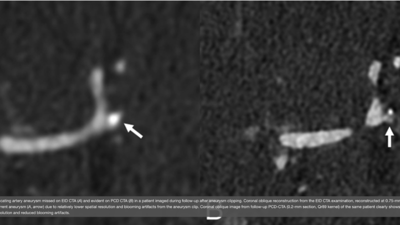
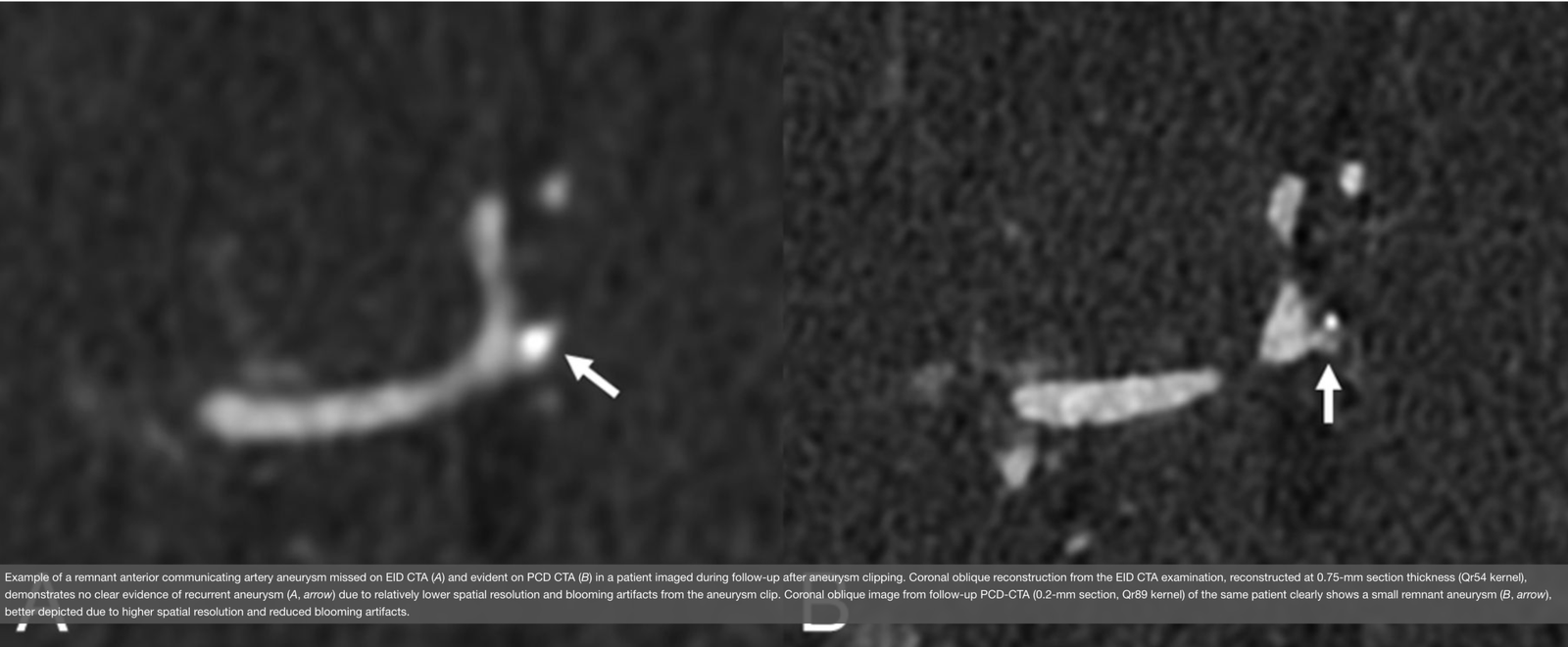
Example of a remnant anterior communicating artery aneurysm missed on EID CTA (A) and evident on PCD CTA (B) in a patient imaged during follow-up after aneurysm clipping. Coronal oblique reconstruction from the EID CTA examination, reconstructed at 0.75-mm section thickness (Qr54 kernel), demonstrates no clear evidence of recurrent aneurysm (A, arrow) due to relatively lower spatial resolution and blooming artifacts from the aneurysm clip. Coronal oblique image from follow-up PCD-CTA (0.2-mm section, Qr89 kernel) of the same patient clearly shows a small remnant aneurysm (B, arrow), better depicted due to higher spatial resolution and reduced blooming artifacts.

Welcome, fellow creatures of the radiology night. Tonight’s subject sits close to my badge and closer to my curiosity: what photon-counting CT can show us inside the skull.
Key takeaways
- Photon-counting CT (PCCT) detectors register individual x-ray photons and sort them by energy, a different physics from conventional energy-integrating detector (EID) CT [1].
- Multiple independent reviews report that PCCT is associated with higher spatial resolution, lower image noise, and higher contrast-to-noise ratio in neuroradiology applications, though most of this evidence comes from narrative reviews and single-center studies [2][3].
- A 2026 single-center, real-world study found PCCT compared favorably with a mix of conventional scanners for image quality and dose in routine neuro exams, though the comparison was pragmatic rather than a controlled test [5].
- Distinguishing gray matter from white matter on standard noncontrast brain CT remains difficult even with PCCT, according to the same literature [3].
The physics
Conventional CT detectors work through an energy-integrating process. Incoming x-rays strike a scintillator, get converted to light, and the detector adds up the total signal from many photons at once. Photon-counting detectors skip that averaging step. They convert each individual x-ray photon directly into an electrical pulse and record its energy as it arrives [1].
This distinction matters for two reasons. Counting photons one at a time removes a source of electronic noise that energy-integrating systems carry along with every measurement. And because each photon’s energy is logged individually, the scanner has spectral information built into every acquisition, with no second pass or extra hardware required [1].
What neuroradiology reviews report
Several independent review groups, working from separate literature, converge on a similar list of PCCT advantages for neuroradiology:
- higher spatial resolution
- reduced noise
- higher contrast between iodine and soft tissue
- lower radiation dose compared with EID-CT [2][7].
These reports span vessel imaging, pituitary adenoma detection, temporal bone imaging, and CT myelography.
One review, synthesizing prior single-center data, reports that PCCT delivers gray-white matter contrast about 15.7% higher than EID-CT, and links this to the possibility of earlier detection of stroke, hemorrhage, and other soft-tissue findings [2].
Dose and contrast-to-noise
A review drawing on dose and contrast-to-noise data found PCCT delivered lower radiation dose than EID-CT (42 ± 17 mGy versus 51 ± 7 mGy) without an apparent drop in overall image quality [3]. The same body of work found that gray-white matter differentiation in the brain remained difficult even on PCCT. [3].
That review also reports that the best contrast-to-noise ratio for gliotic lesions against white matter appeared at virtual monoenergetic reconstructions (images simulating a single chosen x-ray energy) of 60 to 70 keV, while the optimal energy range for hemorrhage varied more widely, from 124 to 164 keV, depending on hemorrhage type [3]. A single default setting will not serve every finding equally, spectral reconstruction has to be tuned to the clinical question.
Small vessels, coils, and metal artifact
Phantom studies on intracranial aneurysms report PCCT spatial resolution up to 110 micrometers in-plane, which the authors suggest could help detect small aneurysms and assess post-treatment findings that are difficult to see on conventional CT [4]. This is still phantom work. The authors themselves note that comprehensive neurovascular reader studies remain scarce [4].
Reduced metal artifact around aneurysm coils and other hardware is a recurring theme in this literature too, tied to the same spectral capabilities that improve soft-tissue contrast elsewhere.
A real-world comparison
An August 2026 single-center study directly compared clinical PCCT against a mix of conventional EID-CT scanners in routine neuroradiology exams, looking at image quality, pathology visibility, and radiation dose [5]. PCCT compared favorably in that comparison. The authors were explicit, though, that this was a pragmatic, real-world comparison rather than a controlled test isolating detector technology as the only variable [5]. Different scanners, different protocols, and clinical variation were all in the mix.
Where the evidence is still thin
A narrative review structured as a SWOT analysis (strengths, weaknesses, opportunities, threats) lays out the barriers alongside the promise: cost, technical complexity, a lack of protocol standardization across sites, and competition from other imaging approaches are all cited as real obstacles to wider neuroradiology adoption [6][7].
A broader, multi-institutional survey adds useful context on top of that. Photon-counting adoption, as distinct from dual-energy CT, is still nascent, with installations concentrated from 2024 onward and many sites lacking dedicated quality control programs [8]. That is a useful corrective against any impression that photon-counting neuroradiology has already settled into routine practice.
None of the sources behind this piece are multicenter trials or meta-analyses. Most of the detailed image-quality data comes from a single vendor’s clinical platform, so how well these findings generalize across manufacturers is not yet established [2][3][5].
The honest summary: the physics is well understood, the early image-quality signal is consistent across independent groups, and the leap from image quality to changed diagnoses or changed outcomes has not yet been measured.
FAQ
What is photon-counting CT (PCCT)?
PCCT is a CT technology in which the detector registers each individual x-ray photon and measures its energy, rather than adding up the total signal from many photons at once as conventional detectors do [1].
Is photon-counting CT better than conventional CT for brain imaging?
For some specific tasks, such as fine neurovascular detail, temporal bone imaging, and imaging around aneurysm coils, current evidence points to advantages in image quality and dose [2][3][4]. For routine noncontrast head CT, gray-white matter differentiation remains a documented weakness even with PCCT, so it is not established as superior across the board [3].
Does PCCT reduce radiation dose in neuroradiology?
A single-center study reported lower dose with PCCT compared with EID-CT, without an apparent loss of image quality [3]. This has not yet been confirmed in multicenter work.
Can PCCT diagnose stroke or hemorrhage more accurately than conventional CT?
No study behind this article measured diagnostic accuracy or clinical outcomes directly. The available evidence describes image-quality metrics, such as contrast-to-noise ratio at specific energy levels, not confirmed changes in diagnosis or patient management [3].
Which scanners does this evidence come from?
Most of the detailed neuroradiology image-quality data in this literature comes from one vendor’s clinical photon-counting platform. Cross-vendor generalizability has not been established from the sources reviewed here [2][3][5].

I have counted photons, coils, and now keV windows. Four hundred years in, and the counting still surprises me.
If you would like the next dispatch from the reading room delivered before the rest of the internet finds it, join our Count Photon Newsletter.
Educational content, not medical advice. Count Photon explains imaging technology and published research for educational purposes. Nothing here is medical advice, and it should never replace a conversation with a qualified healthcare professional about your own care.
References
1. Douek PC, Boccalini S, Oei EHG, Cormode DP, Pourmorteza A, Boussel L, Si-Mohamed SA, Budde RPJ. Clinical Applications of Photon-counting CT: A Review of Pioneer Studies and a Glimpse into the Future. Radiology. 2023.
2. Perera Molligoda Arachchige AS, et al. Neuroimaging with photon-counting computed tomography: A review of clinical applications. World Journal of Radiology. 2025;17(11):113701.
3. Photon-counting CT: An updated review of clinical results. European Journal of Radiology. 2025.
4. Optimizing Photon-Counting Detector CT for Imaging Intracranial Aneurysms. American Journal of Neuroradiology. 2024.
5. Szum A, Moberg F, Kalarakis G, Sadus T, Hashim F, Lilja A, Lundberg J, Granberg T. Clinical Photon-Counting CT in Neuroradiology: A Real-World Paired Comparison with Conventional Energy-Integrating CT. American Journal of Neuroradiology. 2026.
6. Martín-Noguerol T, Santos-Armentia E, Zhu D, Diehn FE, Amrhein TJ, Luna A. Photon-counting CT in neuroradiology: a comprehensive SWOT analysis. European Radiology. 2025.
7. Neuro applications of photon-counting detector CT. British Journal of Radiology. 2025.
8. Spectral CT in practice: insights from an International Atomic Energy Agency survey.