
Figure
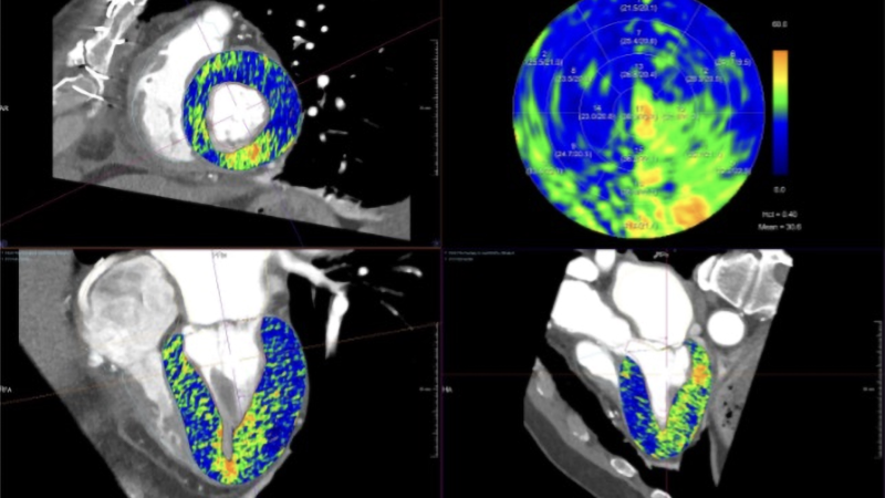
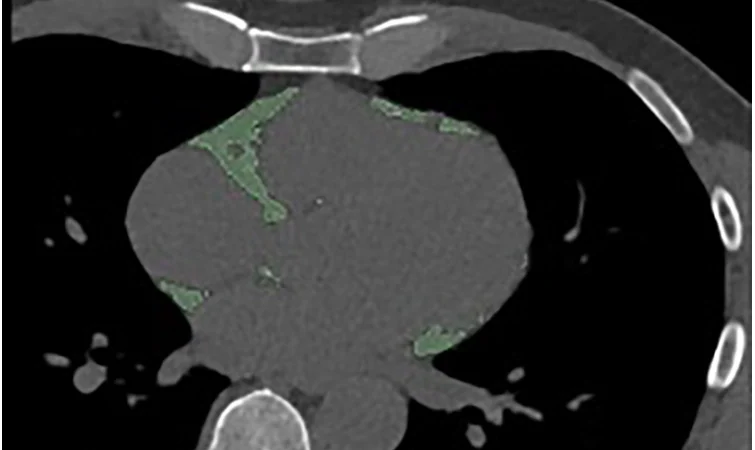
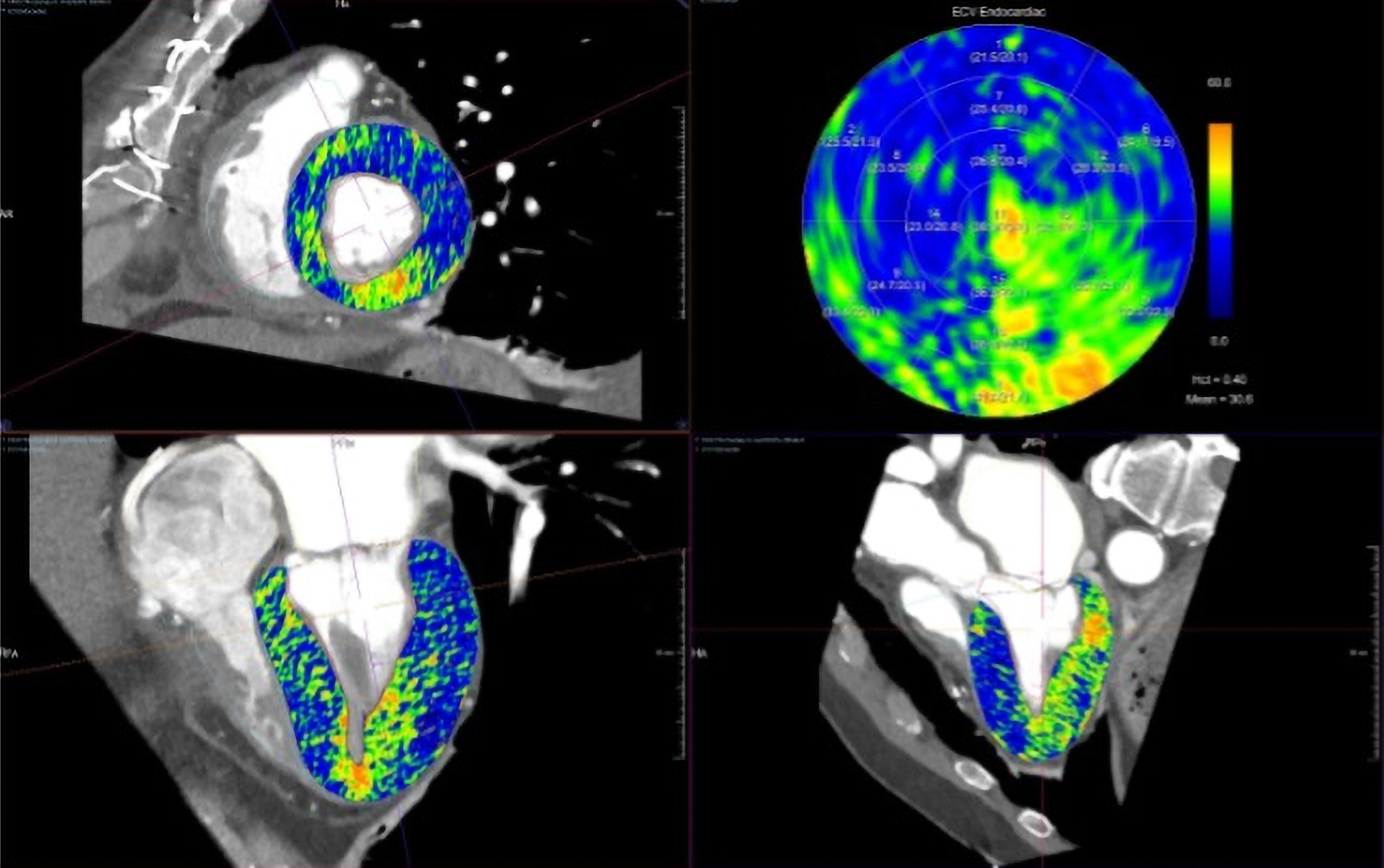
Focal myocardial fibrosis assessed by CT-derived extracellular volume (ECV). A patient with a history of coronary artery disease and prior coronary artery bypass grafting underwent CT coronary angiography for investigation of chest pain. CT-derived ECV mapping demonstrates focally elevated ECV confined to the inferior wall, corresponding to a previous inferior myocardial infarction and consistent with focal replacement fibrosis. https://academic.oup.com/ehjimp/article/4/3/qyag113/8724411

Welcome, fellow creatures of the radiology night. Tonight we count something invisible: the space between heart muscle cells, and whether photon-counting CT can measure it well enough to matter.
Key takeaways
- A 2026 clinical perspective in European Heart Journal Imaging Methods and Practice outlines how photon-counting CT (PCCT) might streamline measurement of cardiac extracellular volume (ECV), a marker linked to myocardial fibrosis and amyloid infiltration [1].
- Small single-center PCCT studies (29–32 patients each, all on the same vendor’s scanner) report strong correlation between PCCT-derived ECV and CMR-derived ECV [1][2][3].
- A separate meta-analysis, not specific to PCCT, found elevated CT-ECV was associated with significantly higher rates of cardiovascular events, mortality, and heart failure hospitalization after aortic valve replacement [4].
- The review’s authors call for standardized protocols and multicenter, prospective outcome studies before PCCT-ECV enters routine clinical workflows [1].
What extracellular volume measures
Extracellular volume is the fraction of heart muscle occupied by the space between cells rather than the cells themselves. Healthy myocardium keeps that fraction low. Scarring from fibrosis, or the abnormal protein deposits of cardiac amyloidosis, expand it [1].
Cardiac MRI (CMR) has long been the reference method for measuring ECV, using contrast timing before and after gadolinium injection. Cardiac CT can estimate the same quantity, and interest in doing so opportunistically, during a coronary CT angiogram or a transcatheter aortic valve implantation (TAVI) planning scan, has grown for a simple reason: many patients already get that scan anyway [1].
How photon-counting CT could simplify the workflow
Conventional CT-ECV methods typically need a non-contrast baseline scan plus a delayed contrast-enhanced scan, then subtraction of the two. PCCT’s inherent spectral sensitivity lets it generate iodine concentration maps directly from a single acquisition, without a dedicated non-contrast pass, according to the review’s authors [1].
Every piece of PCCT-specific evidence discussed in the review comes from the same commercial platform, the Siemens Healthineers NAEOTOM Alpha dual-source scanner, the only clinically available PCCT system in the cited literature [1]. That is worth keeping in mind before generalizing to “photon-counting CT” as a category.
Current PCCT-specific studies
In a study of 29 patients referred for cardiac MRI, PCCT-derived global ECV correlated strongly with CMR-derived ECV (r = 0.91, P < .001), with absolute differences of roughly 2–3%. Dual-energy PCCT acquisition also used about 40% less radiation dose than the single-energy comparison protocol in that same study [2].
In 30 patients with severe aortic stenosis undergoing PCCT for TAVI planning, iodine-map-based ECV agreed closely with a conventional single-energy subtraction method (r = 0.87, mean error 0.9%, limits of agreement −3.3% to 5.0%) [3]. This study did not compare its results against CMR, so its accuracy claims rest on agreement with another CT method rather than the reference standard [3].
Disease-specific single-center studies summarized in the review report similarly strong correlations: in acute myocarditis (32 patients), PCCT-ECV correlated with CMR at basal (r = 0.95) and mid-ventricular (r = 0.91) levels, with a diagnostic threshold near 26.9% and an area under the curve around 0.95; a hypertrophic cardiomyopathy cohort (30 patients) showed comparable agreement [1].
The strongest evidence isn’t PCCT-specific
Two pieces of evidence in the review carry more statistical weight than any single PCCT study, and neither is PCCT-specific.
A meta-analysis of 13 studies and 383 patients, spanning single-energy, dual-energy, and photon-counting CT-ECV methods, reported an excellent overall correlation with CMR-derived ECV (mean r = 0.90, 95% CI: 0.86–0.95), though study quality was described as limited and protocols heterogeneous [1]. That result supports the general concept of CT-ECV, not any particular PCCT advantage over other CT approaches.
Separately, a systematic review and meta-analysis of 10 studies covering 1,223 patients with severe aortic stenosis undergoing valve replacement found elevated CT-ECV was associated with significantly higher rates of cardiovascular events (43.4% vs. 14.0%, OR 4.3, 95% CI 3.19–5.76), all-cause mortality (29.3% vs. 11.6%, OR 3.5, 95% CI 2.28–5.31), and heart failure hospitalization (25.5% vs. 5.9%, OR 4.9, 95% CI 2.28–10.38) over roughly 18 months of follow-up [4]. This is genuinely useful prognostic signal for CT-ECV as a concept. It is not evidence that PCCT-derived ECV specifically predicts outcomes, because none of the studies in that meta-analysis were PCCT studies [4].
Dose and standardization
Early PCCT dose figures look encouraging but remain protocol-dependent. The review cites 2.07 ± 1.9 mSv in one PCCT study and 1.2 mSv (range 0.97–1.75) in another, compared with 1.56–1.98 mSv for single-energy protocols and 1.89–4.8 mSv for dual-energy or spectral protocols elsewhere in the literature [1]. The range tells you protocol choice still matters more than the detector technology alone.
Pooled ECV values also vary by disease state, per a meta-analysis cited in the review: 27.6% (95% CI 25.7–29.4%) in healthy participants, 31.2% in severe aortic stenosis, 50% in cardiac amyloidosis, and 37% in dilated cardiomyopathy. Meta-regression suggested that protocol differences, delayed-imaging timing, single- versus dual-energy technique, and hematocrit method, explained roughly a quarter of the variation between studies [1]. A quarter of the noise traced to methodology alone is a real problem for anyone trying to set a single diagnostic threshold.
Synthetic hematocrit, estimating blood hematocrit from CT attenuation instead of a blood draw, would remove a practical workflow barrier, but the review notes it still needs wider validation before it can be relied upon [1].
What this means for practice today
The review’s authors are explicit about where things stand. They call for consensus guidance and prospective multicenter outcome studies before PCCT-ECV protocols and thresholds can be standardized [1]. Until that happens, cardiac PCCT-ECV remains a promising research and opportunistic-screening tool rather than a validated diagnostic pathway. It cannot replace cardiac MRI for tissue characterization, and the outcome data linking elevated ECV to worse prognosis after valve replacement belongs to CT-ECV broadly, not to PCCT specifically [1][4].
FAQ
What is extracellular volume (ECV) on cardiac CT?
ECV is a measurement of the fraction of heart tissue occupied by the space between cells, which expands with fibrosis or amyloid buildup. It can be estimated from contrast-enhanced CT or cardiac MRI scans [1].
Can photon-counting CT replace cardiac MRI for detecting heart fibrosis?
No. Current evidence shows PCCT-derived ECV correlates with CMR-derived ECV in small studies, but correlation does not establish that the two are interchangeable, and the review does not claim PCCT can substitute for CMR [1][2][3].
Does PCCT-derived ECV predict patient outcomes?
Not based on the evidence in this review. The meta-analysis linking elevated ECV to worse cardiovascular outcomes after aortic valve replacement covers CT-ECV broadly, not PCCT-derived measurements specifically [4].
Is cardiac PCCT-ECV ready for routine clinical use?
Not yet, according to the review’s own authors, who call for standardized protocols and larger multicenter, prospective studies before broader adoption [1].

I have been staring at the inside of my coffin for four centuries. I can wait for a multicenter trial.
If tonight’s arithmetic intrigued you, join the newsletter for more evidence-checked dispatches from the reading room.
Educational content, not medical advice. Count Photon explains imaging technology and published research for educational purposes. Nothing here is medical advice, and it should never replace a conversation with a qualified healthcare professional about your own care.
References
1. Gray R, Rauseo E, Herrey A, Cheasty E, Treibel TA, Pugliese F. Extracellular volume fraction by cardiac computed tomography (ECVCT): opportunities and challenges for clinical implementation. Eur Heart J Imaging Methods Pract. 2026;4(3):qyag113.
2. Aquino GJ, O’Doherty J, Schoepf UJ, et al. Myocardial Characterization with Extracellular Volume Mapping with a First-Generation Photon-counting Detector CT with MRI Reference. Radiology. 2023;307:e222030.
3. Mergen V, Sartoretti T, Klotz E, et al. Extracellular Volume Quantification With Cardiac Late Enhancement Scanning Using Dual-Source Photon-Counting Detector CT. Invest Radiol. 2022;57:406-411.
4. Faggiano A, Gherbesi E, Carugo S, et al. Prognostic value of myocardial computed tomography–derived extracellular volume in severe aortic stenosis requiring aortic valve replacement: a systematic review and meta-analysis. Eur Heart J Cardiovasc Imaging. 2025;26(3):518-531.