
Image Reference
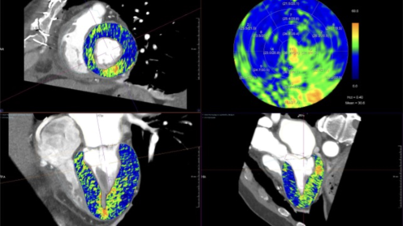
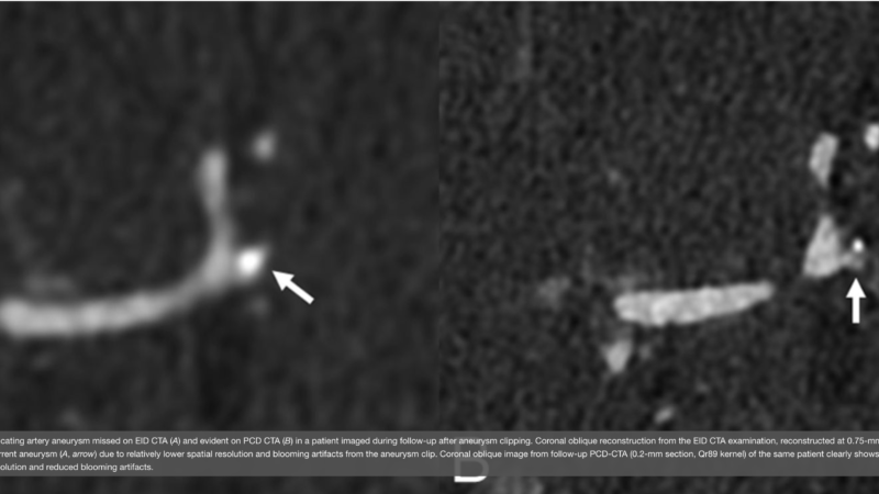
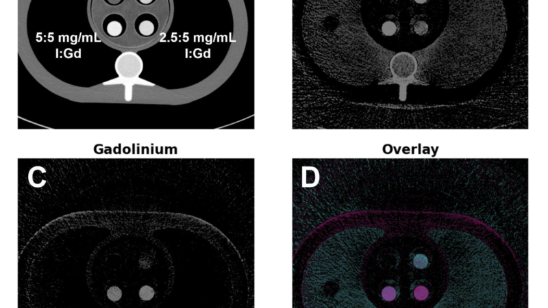
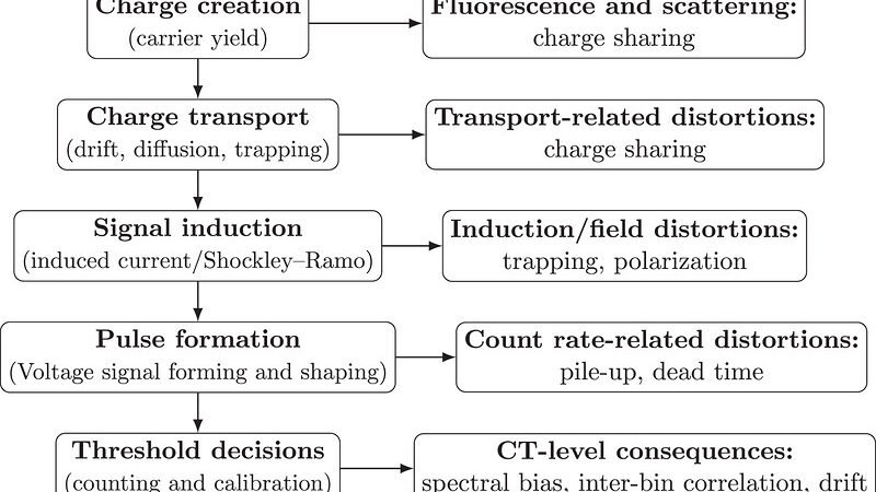
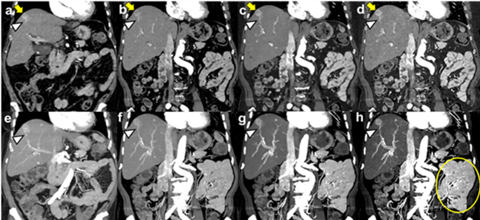
Clinical application of ultra-high-resolution (UHR) photon-counting detector CT (PCD-CT) for visualization of hepatocellular carcinoma (HCC) and tumor-feeding vessels. An 80-year-old female patient with HCV-related liver cirrhosis. a Coronal image obtained 1.5 months earlier using a conventional EID-CT system (Aquilion ONE, Canon Medical Systems), reconstructed at 3-mm slice thickness from 0.5-mm acquisition data. b–d Coronal images obtained using a PCD-CT system from the same 0.2-mm UHR acquisition data with different virtual monoenergetic image (VMI) and reconstruction settings: (b) 70-keV VMI reconstructed at 1 mm using Bv36 and QIR level 2; (c) 40-keV VMI reconstructed at 1 mm using Bv36 and QIR level 2; (d) 40-keV VMI reconstructed at 0.4 mm using a sharp kernel (Bv56) and QIR level 4. e–h Corresponding 15-mm coronal maximum intensity projection (MIP) images. The HCC (yellow arrows) and tumor-feeding artery (white arrowheads) are identifiable on EID-CT images (a, e). PCD-CT images with balanced reconstruction settings (b, f) provide improved visualization of both the tumor and feeding vessels. Lower-keV VMIs further enhance lesion conspicuity and vascular contrast (c, g), which in clinical practice may contribute more substantially than UHR alone. More aggressive UHR reconstruction using thinner slices, a sharp kernel, and higher QIR setting (d, h) provides the clearest depiction of fine vascular structures, including vasa recta and pulmonary vessels (yellow circle in h). However, these reconstructions also increase image noise and may produce overshoot artifacts in high-attenuation regions, resulting in heterogeneous liver parenchymal appearance and reduced overall image quality for routine interpretation. In contrast, MIP images are less susceptible to noise and may particularly benefit from thin-slice UHR reconstruction with sharp kernels.

Welcome, fellow creatures of the radiology night. Tonight’s dossier comes from Tokyo, and for once the counting compulsion in the room isn’t mine — it belongs to the scanner.
Key takeaways
- A newly published narrative review in Abdominal Radiology synthesizes existing literature on photon-counting CT (PCCT) in abdominal imaging, it reports no new patient data of its own [1].
- PCCT detectors count and measure the energy of individual X-ray photons rather than integrating total charge, which lets one acquisition yield multiple spectral reconstructions [1].
- Reported benefits: sharper lesion conspicuity at low keV, better tissue characterization, and potential contrast dose reduction. These are drawn from a still mostly single-center evidence base [1][4].
- A separate multi-institutional expert consensus has proposed standardized protocol settings, such as 70-keV virtual monoenergetic imaging (VMI) for portal venous and pancreas-phase interpretation [5].
What “photon-counting” actually changes about the scan
Conventional CT detectors are energy-integrating detectors (EID): they convert incoming X-rays to light, then to an electrical signal proportional to the total energy absorbed, without distinguishing one photon from another. Photon-counting detector CT (PCD-CT) does something different, it directly converts each X-ray photon into an electrical signal and measures that photon’s individual energy [1].
Because the detector itself is sorting photons by energy, spectral information is built into a standard acquisition rather than requiring a dedicated dual-energy protocol. That’s what enables retrospective reconstructions like virtual monoenergetic imaging (VMI), simulating how an image would look at a single chosen energy level, and material decomposition, which separates an image into maps of substances like iodine or calcium, all from one scan [1].
What the review reports and what kind of evidence that is
It’s worth being precise about what this piece of literature actually is. Despite some secondary coverage describing it as a meta-analysis, the review is in my view a narrative literature review, PubMed lists it simply as “Review,” with no described search protocol and no statistical pooling of outcomes. It was authored by clinicians at a single Japanese academic center, the Institute of Science Tokyo, drawing on 63 prior publications [1].
The review organizes prior findings into four broad categories:
- lesion conspicuity
- tissue characterization
- contrast dose reduction
- quantitative biomarkers
Lesion conspicuity and tissue characterization
On detection, the review states that low-keV imaging improves lesion conspicuity, citing prior work on subtle findings including early-stage pancreatic cancer, small metastases, and inflammatory changes [1]. On characterization, it reports that spectral imaging helps differentiate true enhancement from pseudoenhancement, which the authors say supports more accurate oncologic staging [1].
Both claims are the review’s synthesis of prior single-center and phantom studies, not new comparative data generated for this article [1].
The contrast-dose question
Here the review is notably careful with its own language: it states that amplifying iodine contrast may enable substantial contrast dose reduction while maintaining diagnostic quality, potentially benefiting selected patients who need contrast optimization [1].
How much of this is backed by outcomes data?
Some of the underlying literature does offer direct comparisons. In a single-center study of 66 patients undergoing routine abdominal CT, general image quality on a Likert scale was rated higher for photon-counting CT than for conventional CT (4.74 ± 0.46 vs. 4.25 ± 0.54; p < 0.001) [4].
But even basic protocol questions remain contested. A separate 2024 review in the same journal noted that 40-keV reconstructions produced the best contrast-to-noise ratio for abdominal organs and hypovascular liver metastases, while overall image quality tended to be rated higher at 50 and 60 keV compared with conventional CT, and lower at 40 keV [2]. In other words, radiology colleagues studying this question haven’t converged on one “correct” energy level yet.
This review acknowledges its own field’s growing pains directly, noting that challenges including increased data volume, technical limitations, cost-effectiveness, and the need for optimized reconstruction strategies remain despite PCCT’s advantages [1].
Toward standardization: an expert consensus
Separately from this review, a multi-institutional group, the Society of Abdominal Radiology’s Photon-Counting Detector CT Emerging Technology Commission, published a consensus statement the same year. Nine radiologists from nine institutions used a structured consensus-minus-one method and reached agreement on 20 protocol features, including that 70-keV VMI is recommended for primary interpretation of portal venous and multiphase pancreas PCCT, and that low virtual monoenergetic levels should be viewed for multiphase aortic CTA [5].
The bottom line for now
PCCT changes abdominal CT protocols primarily by adding spectral flexibility after a single acquisition, radiologists can choose or reconstruct different virtual energy levels and material maps from one scan, rather than needing a dedicated dual-energy acquisition. Whether that flexibility reliably translates into better diagnosis, staging, or patient outcomes is still being worked out, largely through single-center studies, phantom experiments, and expert consensus rather than large trials [1][2][4][5].
But then, what about spectral-detector CT?
Dual-layer spectral-detector systems such as Philips’ IQon and newer Spectral CT 7500/Verida platforms deliver a similar core promise: every acquisition is inherently spectral, so radiologists can generate virtual monoenergetic images, material decomposition maps, and effective-Z data retrospectively, without needing to plan for dual-energy mode in advance. In that sense, the two technologies are solving the same clinical problem, retrospective spectral flexibility from a single scan, through different hardware approaches: energy discrimination at the detector layer versus photon-by-photon energy discrimination at the source.
Dual-layer spectral CT, by contrast, has a longer clinical track record, more published outcome data across abdominal and vascular applications, and because it doesn’t require new detector materials or acquisition workflows is generally positioned as a lower-cost, easier-to-integrate upgrade path for institutions already invested in conventional CT infrastructure. [6][7][8][9][10][11][12]
Whether PCCT’s theoretical resolution and noise advantages translate into meaningfully better diagnostic accuracy or outcomes compared to mature dual-layer systems, as opposed to simply offering a different route to similar spectral information, remains an open question.
FAQ
Does photon-counting CT require an entirely different abdominal protocol than conventional CT?
Not entirely — the acquisition itself is broadly similar, but the spectral data it captures opens up reconstruction choices (like which virtual energy level to view) that conventional CT doesn’t offer from a single scan [1].
What is virtual monoenergetic imaging (VMI), and why does it matter here?
VMI simulates how an image would look at a single chosen X-ray energy level. Different keV levels can favor different tasks, for example, low keV for lesion conspicuity versus higher keV for overall image quality, which is part of why standardization efforts are underway [1][2][5].
Does PCCT reduce contrast dose in abdominal imaging?
The review describes this as a possibility for selected patients under certain conditions, not an established general benefit, the underlying evidence is still limited [1].
Is photon-counting CT now the standard of care for abdominal imaging?
No. The evidence discussed here comes from a narrative review, single-center studies, and an expert consensus survey, none of which establishes PCCT as a replacement for conventional CT in routine abdominal practice [1][4][5].
Why do sources disagree on the best keV setting?
Because the underlying studies are still small and vary in what they’re optimizing for, contrast-to-noise ratio versus overall subjective image quality and the field hasn’t yet settled this with large comparative data [2].
References
1. Yokoyama K, Kawasaki Y, Fujii M, et al. “Seeing the invisible: practical strategies to maximize the clinical impact of photon-counting CT in abdominal imaging.” *Abdominal Radiology*, 2026 (online ahead of print).
2. Onishi H, Tsuboyama T, Nakamoto A, et al. “Photon-counting CT: technical features and clinical impact on abdominal imaging.” *Abdominal Radiology*, 2024;49(12):4383-4399.
4. “Initial experience on abdominal photon-counting computed tomography in clinical routine: general image quality and dose exposure.” 2023 (n=66, single-center retrospective study).
5. Dane B, Ananthakrishnan L, Marin D, et al. “Adult Abdominal Photon-Counting CT Protocols: A Multiinstitutional Consensus of the Society of Abdominal Radiology Photon-Counting Detector CT Emerging Technology Commission.” AJR, 2025;225:e2533625.
6. Rassouli N, Etesami M, Dhanantwari A, Rajiah P. Detector-based spectral CT with a novel dual-layer technology: principles and applications. Insights Imaging. 2017;8(6):589-598.
7. McCollough CH, Leng S, Yu L, Fletcher JG. Dual and multienergy CT: principles, technical approaches, and clinical applications. Radiology. 2015;276(3):637-653.
8. Wang W, et al. The clinical applications of dual-layer spectral detector CT in digestive system diseases. Eur Radiol. 2024.
9. Dual-layer spectral detector CT for contrast agent concentration, dose and injection rate reduction: Utility in imaging of the superior mesenteric artery. Eur J Radiol (ScienceDirect). 2022.
10. Rajiah PS, Kambadakone A, Ananthakrishnan L, Sutphin P, Kalva SP. Vascular Applications of Dual-Energy Computed Tomography. Radiol Clin North Am. 2023;61(6):1011-1029.
11. Cao W, et al. Dual-Head Pix2Pix Network for Material Decomposition of Conventional CT Projections with Photon-Counting Guidance. 2025.
12. Sartoretti T, et al. Photon Counting CT: Clinical Applications and Future Developments. PMC. 2021;8409241.

That’s tonight’s ledger, dear colleagues — carefully counted, nothing rounded up. I look forward to the day a genuine multicenter trial lets me update these numbers with real enthusiasm rather than careful caveats.
Fancy more nightly dispatches like this one? Join the Count Photon newsletter — the Count promises no exaggeration, only citations.
Educational content, not medical advice. Count Photon explains imaging technology and published research for educational purposes. Nothing here is medical advice, and it should never replace a conversation with a qualified healthcare professional about your own care.