
Image credentials
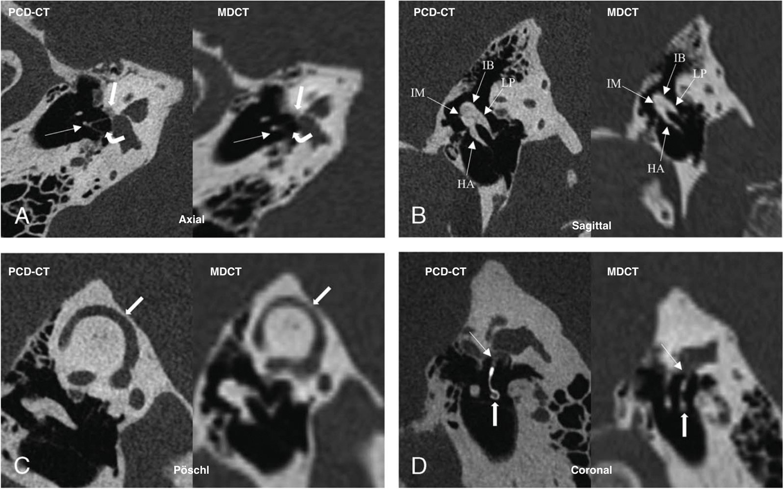
Figure 1.. PCD-CT and MDCT Comparison
Representative images comparing the PCD-CT scan of a temporal bone (left) to corresponding images from a 64-slice multi-detector computed tomography (MDCT) temporal bone scan obtained in the community (right) in a patient with otosclerosis. A–Patient’s right temporal bone. A, Oblique axial reconstructions in the plane of the long axis of the stapes. Note the difference in the resolution of the stapes superstructure (thin arrows), intercrural space (curved thick arrows), and the focus of otosclerosis at the fissula ante fenestra (straight thick arrows). B, Oblique sagittal reconstructions in the molar tooth plane. Again, note the difference in resolution demonstrating the ossicular anatomy (HA, handle of the malleus; IM, incudomalleolar joint; IB, incus body; LP, long process of incus). C, Oblique sagittal reconstructions in the Pöschl plane. Note the clear delineation between the superior semicircular canal and the middle fossa (thick arrows) afforded by the PCD-CT compared with the MDCT. D, Oblique coronal reconstructions of the left temporal bone along the long axis of the stapes prosthesis (thick arrows), with the shape of the prothesis much more clearly defined on PCD-CT. The prosthesis is displaced from the stapes footplate, with some scarring or debris separating the distal prosthesis from the oval window (thin arrows), much better demonstrated on PCD-CT. Note, labels have been added to the original figure for clarity. Source: Macielak RJ, Benson JC, Lane JI, et al. Photon-counting detector CT for temporal bone imaging: Up to three times the resolution at half the radiation dose. Otol Neurotol. 2022 Dec 1;43(10):e1205–7.

Welcome, fellow creatures of the radiology night. Tonight we descend into the temporal bone, a small patch of skull that hides three of the tiniest bones in the human body, and ask whether photon-counting CT’s famous resolution actually helps anyone see them better.
Key takeaways
- A 2026 review by a Mayo Clinic group finds that photon-counting CT (PCCT) consistently shows finer detail in temporal bone structures than conventional CT [1].
- Several small, single-center studies report better visualization scores for ossicular prostheses, cochlear implant electrodes, and related structures, often at lower radiation dose [2][3][4].
- The review’s own conclusion: evidence that this translates into better diagnosis or surgical outcomes “remains limited” [1].
- Ultra-high-resolution modes can reach well under 0.2 mm, but the sharpest settings add noise, so clinical protocols typically use smoother reconstruction kernels [8].
Why temporal bone imaging demands fine detail
The temporal bone houses the ossicles (malleus, incus, and stapes), along with the cochlea and semicircular canals of the inner ear. These structures measure fractions of a millimeter. A prosthesis joint gap, a subtle erosion, or a thin spot over the superior semicircular canal (SSCD) can separate a straightforward diagnosis from a missed one. Conventional energy-integrating detector CT (EID-CT) has long been the workhorse here, but its spatial resolution runs up against real physical limits when the structures in question are this small.
What the new review states
A 2026 narrative review from a Mayo Clinic group, Epperson, Leng, Lane, and Carlson, surveyed recent literature on photon-counting detector CT (PCD-CT) for temporal bone imaging [1]. This is a review of other groups’ work, not a new study of its own, and its authors are careful about the line between what has been shown and what has not.
Their summary: PCD-CT gives substantially better visualization of submillimeter temporal bone structures than conventional CT [1]. That part of the picture is consistent across the studies they surveyed. What is not yet established, in their own words, is whether seeing these structures more clearly changes what a diagnosis or a surgical plan actually looks like.
The evidence behind the visualization gains
The studies underlying that conclusion are consistent, independent, and worth naming.
In a foundational Mayo Clinic study, an investigational PCD-CT system with spatial resolution of 150 micrometers or better produced superior visualization of critical temporal bone structures at an average 31% lower radiation dose than conventional CT, in the same patients [2].
A separate single-center comparison against a Siemens Somatom Force multidetector scanner found the odds of better structure visualization were 354 times higher with PCD-CT (95% CI, 75-1673; p<0.0001), alongside a lower dose-length product: 74 mGy·cm versus 95 mGy·cm [3].
A small intra-patient study (seven patients) found PCD-CT gave better visualization than conventional CT for all six evaluated middle-ear structures assessed, including the incudostapedial and incudomalleolar joints, each at p<0.05 [4]. A related review reports noise-matched dose reductions approaching 80% when PCD-CT and conventional CT images are compared directly [5].
These are small, single-center numbers, several sharing overlapping authorship, and none of them measure diagnostic accuracy against a surgical or pathological reference standard. The direction of the finding, better visualization at equal or lower dose, repeats across independent groups.
The resolution-noise trade-off
Here is where the “0.2 mm” figure often quoted for PCCT needs a caveat. A technical paper notes that PCD-CT in-plane resolution can reach 0.125 mm, but that the sharpest reconstruction settings introduce excessive image noise [8]. In practice, clinical temporal bone protocols use smoother kernels rather than the maximum resolution the hardware can produce. The number on the spec sheet and the number a radiologist actually reads off the console are not automatically the same thing.
What’s still missing
None of the studies reviewed measured whether better visualization changes management: whether a surgeon operates differently, whether cochlear implant candidacy shifts, or whether hearing outcomes improve. The review’s own open questions include exactly this gap. No diagnostic-accuracy study against a surgical or histopathologic reference standard, and no clinical-outcomes study, has yet been published for PCD-CT in temporal bone imaging [1].
Readers may notice that cholesteatoma, a common reason for ordering temporal bone CT, doesn’t appear in the findings above. The primary review’s available findings concern ossicular prostheses, cochlear implant electrodes, and SSCD specifically. Cholesteatoma detection with PCD-CT simply isn’t addressed in the source available to us. Whether the same resolution gains hold there is an open question.
What this means for practice, today
In short: Photon-counting CT reliably shows finer temporal bone detail at equal or lower dose across multiple independent studies, and whether that changes diagnosis or outcomes is still unproven.
FAQ
What is photon-counting CT and how is it different from conventional CT?
Photon-counting detector CT (PCD-CT) counts individual X-ray photons and sorts them by energy, rather than measuring the combined energy of many photons at once as conventional energy-integrating detector CT (EID-CT) does. This generally allows finer spatial resolution and lower image noise per dose.
Does photon-counting CT improve cholesteatoma detection?
The available evidence on this specific question is limited. The 2026 review covering this topic addresses ossicular prostheses, cochlear implant electrodes, and superior semicircular canal dehiscence, not cholesteatoma specifically, so no claim about cholesteatoma detection can be drawn from it.
Is photon-counting CT proven to change surgical outcomes for ear disease?
No. Every study identified so far measures image quality or visualization scores, not diagnostic accuracy against a reference standard or patient outcomes. That evidence gap is explicitly acknowledged by the review’s own authors [1].
Why doesn’t imaging always use the highest resolution setting available?
Because resolution and noise trade off against each other. The sharpest reconstruction kernels reach very fine detail but introduce more image noise, so clinical protocols typically use a smoother setting balanced for diagnostic use [8].
Are these findings based on large clinical trials?
No. The underlying studies are small and single-center, mostly measuring image quality rather than diagnostic accuracy. No multicenter trial has yet been published on this specific application [1].

Until the next study lands on my desk (I do read quickly; four centuries builds a certain fluency), consider photon-counting CT’s temporal bone story a promising chapter still being written, not a closed case.
Stay curious, and mind the noise floor.
Want more evidence-checked dispatches from the reading room? Subscribe to the Count Photon newsletter.
Educational content, not medical advice. Count Photon explains imaging technology and published research for educational purposes. Nothing here is medical advice, and it should never replace a conversation with a qualified healthcare professional about your own care.
References
1. Epperson MV, Leng S, Lane JI, Carlson ML. Photon-counting detector computed tomography for temporal bone: does higher resolution matter? Curr Opin Otolaryngol Head Neck Surg. 2026 Jul 7 (online ahead of print).
2. Benson JC, Rajendran K, Lane JI, et al. A new frontier in temporal bone imaging: photon-counting detector CT demonstrates superior visualization of critical anatomic structures at reduced radiation dose. AJNR Am J Neuroradiol. 2022;43:579-584.
3. Photon-counting CT allows better visualization of temporal bone structures in comparison with current generation multi-detector CT. Insights Imaging / European Radiology Experimental.
4. Improved visualization of temporal bone structures with photon-counting detector CT: an intra-patient comparison.
5. Benson JC, Campeau NG, Diehn FE, et al. Photon-counting CT in the head and neck: current applications and future prospects. AJNR Am J Neuroradiol. 2024;45:1000-1005.
6. Ultra-High-Resolution Temporal Bone Anatomy Using Photon-counting CT: Added Value of Improved Spatial Resolution. RadioGraphics. 2024.
7. Photon-Counting Detector CT for Temporal Bone Imaging: Up to Three Times the Resolution at Half the Radiation Dose. Otol Neurotol. 2022;43(10):e1205-e1207.
8. Ultra-High-Resolution Photon-Counting-Detector CT with a Dedicated Denoising Convolutional Neural Network for Enhanced Temporal Bone Imaging. AJNR Am J Neuroradiol. 2025 (early online).