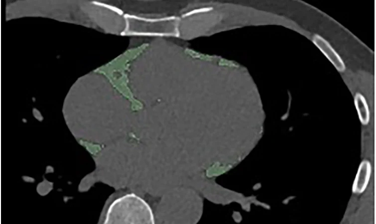

Figure 4a:
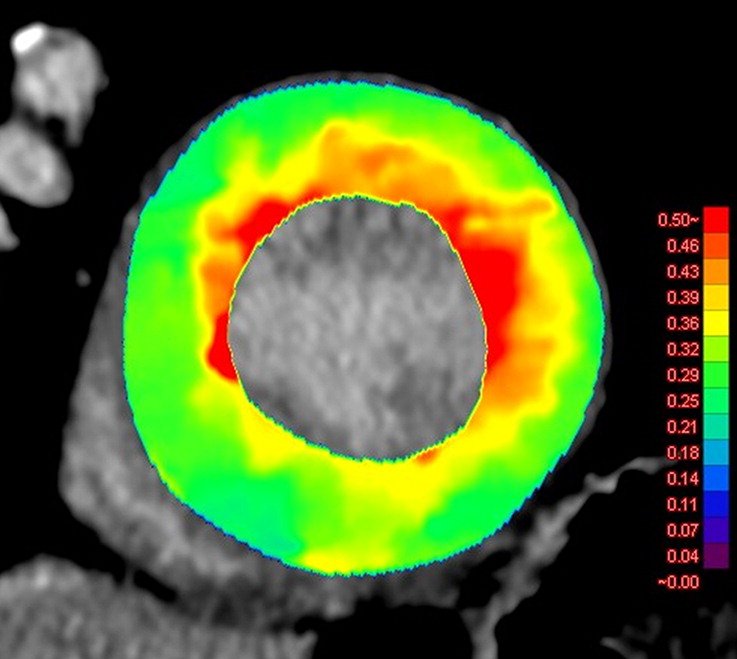
Images show comparison of (a) CT-derived extracellular volume (ECV) and (b) cardiac MRI-derived ECV in a 56-year-old woman with hypertrophic cardiomyopathy. Both CT-derived ECV and cardiac MRI–derived ECV images show significantly elevated myocardial ECV predominantly in subendocardium. CT-derived ECV and cardiac MRI–derived ECV quantifications were comparable. https://pubs.rsna.org/doi/10.1148/ryct.2019180003

Welcome, fellow creatures of the radiology night. Tonight’s subject is iodine, and how photon-counting CT (PCCT) might be persuaded to see it more clearly, several minutes after it has already left the building.
Key Take-Aways
- Phantom research already confirms that energy threshold selection and iterative reconstruction settings measurably affect iodine contrast-to-noise ratio (CNR) on PCCT [2][3].
- A new single-center report (Schramm et al.) reportedly tested these same parameters for delayed, or “late” iodine enhancement imaging.
- Angiography and delayed-enhancement protocols on PCCT remain a work in progress across vendors and sites, not a settled standard.
How energy thresholds and reconstruction shape iodine images
A photon-counting CT (PCCT) detector registers each individual X-ray photon and sorts it into an energy bin, rather than summing total energy the way a conventional energy-integrating detector (EID) does. The threshold that defines each bin, the minimum photon energy counted before a photon is assigned to that bin, shapes the spectral data that comes out the other end.
That matters for iodine because iodine has a K-edge, a jump in X-ray absorption at a specific energy, that makes it especially sensitive to which photons get counted where. Reconstruction choices add a second layer: iterative reconstruction reduces image noise computationally, and virtual monoenergetic images (VMI) let a scan be reconstructed as if acquired at a single chosen energy, tuned to make iodine stand out.
Delayed, or “late” iodine enhancement imaging adds a third variable: timing. These scans are taken minutes after contrast injection, once iodine concentration in tissue has already dropped from its first-pass peak, which makes contrast-to-noise ratio (CNR) harder to win in the first place.
What phantom studies have already shown
Two verified phantom studies give this some grounding. Sawall and colleagues tested a whole-body PCCT system and found that threshold selection measurably changes iodine image contrast and spectral separation [2]. Booij and colleagues, working with an anthropomorphic phantom, found that iterative reconstruction strength and VMI energy level both independently affect iodine CNR when comparing photon-counting detector CT to dual-source energy-integrating CT [3].
Both are phantom work, useful for establishing that a physical effect exists and roughly how large it is under controlled conditions, not for telling a technologist what to dial in for a real patient.
A new single-center report
A study by Schramm and colleagues, indexed on PubMed under PMID 42440227, tested threshold selection and iterative reconstruction methods to identify PCCT protocols that maximize CNR for delayed enhancement imaging, without a dose penalty [1].
Among 74 patients scanned between May 2024 and July 2025, 41 (55%) showed myocardial scar — mostly transmural, with 59% having an implantable cardioverter defibrillator — and their images were reconstructed with two kernels (Qr40, Qr36) and two slice thicknesses (0.4 mm, 2 mm), then analyzed across monoenergetic levels from 40 to 120 keV.
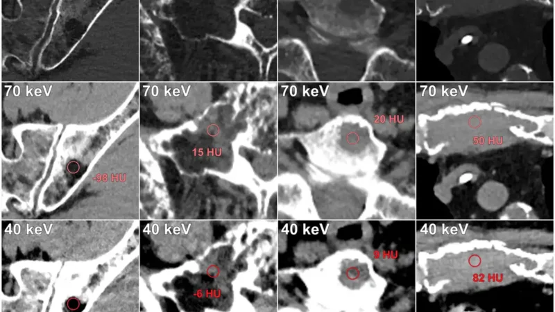
Virtual monoenergetic images at 40 keV consistently yielded the highest CNR and SNR regardless of reconstruction settings, peaking at a median CNR of 5.36 with 2-mm slices and the Qr36 kernel, while scar pattern, ICD presence, and BMI had no significant effect.
In an exploratory subgroup of 16 patients who also underwent late gadolinium enhancement MRI, CNR did not differ significantly between the two modalities (p = 0.178), though the MRI values were numerically higher. The authors conclude that 40-keV VMI with 2-mm slice thickness is the optimal configuration for PCD-CT LIE.
What this means for protocol writers, for now
Systematic threshold and reconstruction optimization could help move late-enhancement and angiography protocols on PCCT away from ad hoc, site-specific settings and toward something more consistent.
Threshold effects have already been shown to depend on the specific PCCT hardware in phantom work, so what optimizes CNR on one vendor’s system may not transfer cleanly to another’s. Angiography and delayed-enhancement imaging on PCCT are still being written, one careful study at a time.
Frequently asked questions
What is late iodine enhancement imaging?
It is a CT scan taken several minutes after contrast injection, used to detect abnormal iodine retention in tissue, such as scarred heart muscle or certain lesions.
Does energy threshold selection actually change image quality on photon-counting CT?
Yes, in phantom studies. Threshold choice on a photon-counting detector measurably affects iodine contrast and spectral separation, and reconstruction settings independently affect contrast-to-noise ratio [2][3]. Whether a specific set of optimal thresholds exists for clinical late-enhancement imaging is what the new single-center report claims to address, though that claim has not yet been independently verified.
Is there a standard PCCT angiography protocol yet?
Not that this dossier can confirm. Protocols for angiography and delayed-enhancement imaging on photon-counting CT remain an active area of technical optimization rather than an established standard.
I shall keep this study on my desk, so to speak, until further review has been done. Four centuries have taught me that a number worth trusting is worth waiting for.
If you would like the update when I have actually read it, the newsletter is the place. Sign up, and I will count you among the first to know.
Educational content, not medical advice. Count Photon explains imaging technology and published research for educational purposes. Nothing here is medical advice, and it should never replace a conversation with a qualified healthcare professional about your own care.
References
1. Schramm et al. Image-Quality Optimization for Late Iodine Enhancement on PCCT. PubMed ID 42440227.
2. Sawall S, Klein L, Wehrse E, et al. Threshold-dependent iodine imaging and spectral separation in a whole-body photon-counting CT system. European Radiology. 2021.
3. Booij R, van der Werf NR, Dijkshoorn ML, van der Lugt A, van Straten M. Assessment of Iodine Contrast-To-Noise Ratio in Virtual Monoenergetic Images Reconstructed from Dual-Source Energy-Integrating CT and Photon-Counting CT Data. Diagnostics. 2022.