Photon-Counting CT Myelography and CSF-Venous Fistulas
Soren K. Christensen, Peter G. Kranz, Ajay A. Madhavan, Michael D. Malinzak, Daphne Zhu, Jay Willhite, Linda Gray and Timothy J. Amrhein
American Journal of Neuroradiology July 2026, ajnr.A9507; DOI: https://doi.org/10.3174/ajnr.A9507
Welcome, fellow creatures of the radiology night. Tonight’s subject is a leak so subtle it can hide from an entire CT scanner, the CSF-venous fistula, and a new single-center study asking whether photon-counting CT myelography helps radiologists feel more certain when they find one.
Key takeaways
A retrospective single-center study of 628 CT myelography exams at Duke University found photon-counting CT myelography (PCD-CTM) produced significantly higher diagnostic certainty scores than conventional energy-integrating CT myelography (EID-CTM) for CSF-venous fistulas (CVFs) [1].
Raw fistula detection was numerically higher with PCD-CTM (69.6%) than EID-CTM (61.0%), but that difference did not reach statistical significance (P = .07) [1].
This is single-center, retrospective, non-randomized evidence from one research group, not a multicenter trial, and not confirmation against a surgical or embolization reference standard.
What the study actually measured
CSF-venous fistulas are a recognized but frequently subtle cause of spontaneous intracranial hypotension (SIH), a condition of low spinal fluid pressure that often produces severe headaches. Finding the fistula on imaging has long been the hard part, hence the interest in higher-resolution CT myelography techniques.
Researchers at Duke University Medical Center retrospectively reviewed 628 CT myelography exams performed in 456 patients between 2020 and 2025 (63.2% women, mean age 58.0 years) [1]. Of these, 148 exams were performed on a photon-counting detector CT scanner and 480 on a conventional energy-integrating detector CT scanner. Group assignment reflected which scanner was available at the time of each exam, not randomization, a distinction that matters for how much weight the comparison can bear.
Two neuroradiologists, with consensus adjudication, scored each exam using the Duke CSF-Venous Fistula Confidence Score (DCCS), a 0–3 structured scale the same group had developed and validated in an earlier single-center study [4]. A score of 2 or higher counted as fistula detection.
I do enjoy a well-built measuring instrument, four centuries of counting grains of rice will do that to a vampire, but it’s worth remembering this scale was built and tested at the same institution running this comparison.
The finding: more confidence, not yet more confirmed fistulas
Across the full cohort, CVFs were detected (DCCS ≥2) in 63.1% of exams (396/628) [1]. The comparison between scanner types produced two distinct results, and the distinction is the whole story here.
First, PCD-CTM showed a statistically significant shift toward higher DCCS certainty scores compared with EID-CTM (P = .006) [1]. On ordinal logistic regression, exams performed with PCD-CTM had 66% greater odds of falling into a higher certainty category than EID-CTM exams (odds ratio 1.66; 95% CI, 1.17–2.36; P = .005) [1]. That’s a solid, statistically robust result — for reader confidence.
Second, raw detection rates, the more clinically important number, since it asks “did we actually find the fistula?” rather than “how sure were we?”, were numerically higher with PCD-CTM (69.6%) than EID-CTM (61.0%), but this difference did not reach statistical significance (P = .07) [1].
In plain terms: this study supports a claim about diagnostic certainty, not (yet) a claim about superior diagnostic accuracy. Those are genuinely different things, and the paper is careful about which one it’s showing.
How this fits the group’s prior work
An earlier, smaller study from largely the same investigators found that PCD-CT myelography scored significantly better on subjective image quality than EID-CT myelography, across three independent readers (all P < .05) [2]. Separately, an independent single-center study at a different institution (57 patients, decubitus positioning) reported PCD-CTM identified definitive fistulas in 56.0%–76.5% of patients depending on pretest probability tier, though that study did not directly compare against EID-CTM [3].
Taken together, these three papers point the same direction: photon-counting detectors appear to help see and describe CSF-venous fistulas with more confidence.
What this study does not show
A few limitations are worth stating plainly, because the abstract itself is candid about them.
Not randomized. The unequal group sizes (148 vs. 480) and the 2020–2025 date range suggest the scanner-type split largely tracks which machine was available over time, not a controlled comparison, leaving room for confounding by era, referral patterns, or case mix.
Certainty, not confirmed accuracy. The DCCS is a structured confidence score assigned by study readers, not confirmation of true fistula presence against an independent reference standard like digital subtraction myelography, surgery, or transvenous embolization.
Blinding not reported. The abstract does not describe whether readers were blinded to scanner type, relevant for an endpoint that is inherently subjective.
Single institution. Everything here comes from one academic medical center, using a confidence scale that center itself developed.
Full text unavailable. This dossier was built from the published abstract; scanner vendor/model, radiation dose, and conflict-of-interest disclosures could not be verified.
Why finer resolution plausibly matters here
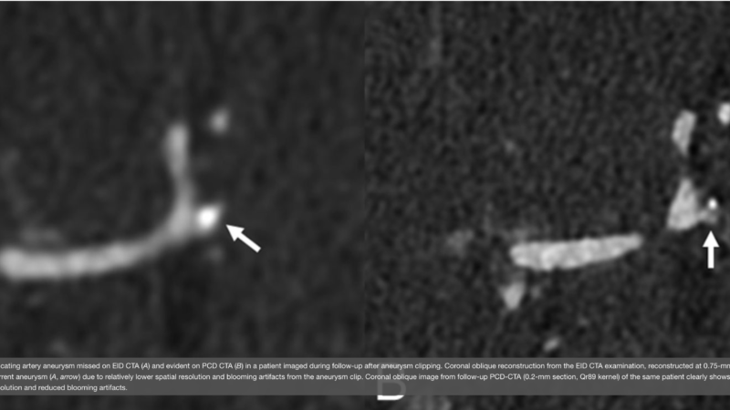
CSF-venous fistulas are often faint, thread-like contrast tracts running alongside the spine, the kind of finding that can vanish into image noise on conventional CT. Photon-counting detectors are built to reduce electronic noise and improve spatial resolution [see our companion explainer on photon-counting CT detector basics], which is a physically reasonable explanation for why readers might feel more confident spotting a subtle leak.
FAQ
Does photon-counting CT find more CSF-venous fistulas than conventional CT?
In this single-center study, raw detection was numerically higher with photon-counting CT (69.6% vs. 61.0%), but the difference was not statistically significant (P = .07) [1]. The study’s strongest, statistically significant finding was about reader certainty.
Is this evidence multicenter or randomized?
No. This is a retrospective, single-center, non-randomized comparison from one Duke research group [1].
What is the Duke CSF-Venous Fistula Confidence Score (DCCS)?
A 0–3 structured scale developed at Duke to standardize how confident a radiologist is that a CSF-venous fistula is present on imaging; a score of 2 or higher was counted as detection in this study [4].
References
1. Christensen SK, Kranz PG, Madhavan AA, et al. Improved Diagnostic Certainty of Photon-Counting CT Myelography Compared with Energy-Integrating CT for CSF-Venous Fistulas in Spontaneous Intracranial Hypotension. AJNR Am J Neuroradiol. 2026 Jul 1 (ahead of print).
2. Schwartz FR, Kranz PG, Malinzak MD, et al. Myelography Using Energy-Integrating Detector CT Versus Photon-Counting Detector CT for Detection of CSF-Venous Fistulas in Patients With Spontaneous Intracranial Hypotension. AJR Am J Roentgenol. 2024;222(4):e2330673.
3. Madhavan AA, Yu L, Brinjikji W, et al. Diagnostic Performance of Decubitus Photon-Counting Detector CT Myelography for the Detection of CSF-Venous Fistulas. AJNR Am J Neuroradiol. 2023 (early edition).
4. Amrhein TJ, Zhu D, Gray L, et al. Reporting the Degree of Certainty of CSF-Venous Fistulas in Patients with Spontaneous Intracranial Hypotension: The Duke CSF-Venous Fistula Confidence Score. AJNR Am J Neuroradiol. 2025;46(11):2399-2405.
Enjoyed this dispatch from the reading room? Join The Count Photon newsletter for weekly, carefully hedged updates on photon-counting CT research.
Educational content, not medical advice.Count Photon explains imaging technology and published research for educational purposes. Nothing here is medical advice, and it should never replace a conversation with a qualified healthcare professional about your own care.