
TL;DR
- GE Healthcare has launched the world’s first clinical trials of photon counting CT technology using revolutionary deep silicon detectors at Karolinska University Hospital, marking a major milestone in medical imaging after 30 years of research and development.
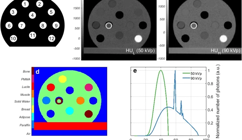
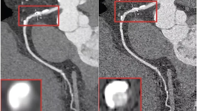
- Unlike traditional CT scanners that use a two-step conversion process, this breakthrough technology directly converts X-ray photons into electrical signals, allowing each photon to be counted individually and its energy level measured, resulting in significantly clearer images with reduced radiation exposure for patients.
- Following GE’s acquisition of Swedish startup Prismatic Sensors in November 2020, the rapid progression to clinical trials in less than a year demonstrates what Stanford professor Norbert Pelc calls “the most significant improvement in CT technology in decades,” with potential applications spanning oncology, cardiology, neurology, and general diagnostics.
GE Healthcare has achieved a significant milestone as its groundbreaking photon counting CT technology begins large-scale clinical trials for the first time. The trials, supervised by the prestigious Karolinska Institutet and MedTechLabs at Karolinska University Hospital, mark a pivotal moment in medical imaging technology.
What sets GE’s technology apart is its use of deep silicon detectors—a world first in photon counting CT systems. This innovative approach promises to unlock the full potential of photon counting technology by delivering exceptional spatial resolution without compromising counting rate or spectral capabilities.
The Technology Behind the Innovation
Traditional CT vs. Photon Counting CT
Traditional CT systems use a two-step process to create images:
- X-rays hit scintillator materials (like gadolinium sulfide or cesium iodide)
- These materials convert X-rays into visible light
- Photoelectric devices then convert this light into electrical signals
This indirect conversion process has limitations—photons scatter in multiple directions, causing signal loss and reduced spatial resolution. Additionally, traditional systems cannot capture individual photon energy levels.
In contrast, photon counting detectors use semiconductor materials to directly convert X-ray photons into electrical signals in a single step. This direct conversion allows:
- Individual photon counting for more accurate imaging
- Energy level quantification for superior spectral information
- Enhanced spatial and spectral resolution
- Reduced radiation exposure
- Improved contrast-to-noise ratios
Why Deep Silicon?
GE’s journey to deep silicon technology spans three decades. After developing the world’s first photon counting CT prototype using cadmium-based detectors in 2006, GE ultimately chose pure silicon as their semiconductor material of choice.
Silicon offers several advantages:
- Exceptional purity
- Abundant availability
- Well-established manufacturing infrastructure
However, silicon’s relatively low atomic number presented a challenge—traditional face-up positioning wouldn’t allow sufficient photon absorption. GE solved this through their patented “Deep Silicon” approach: positioning the silicon sensor on its side to maximize absorption depth. This configuration enables the detector to process hundreds of millions of photons per second, creating clearer images than conventional CT systems.
Comparison with Competitors
While Siemens Medical recently launched the NAEOTOM Alpha using cadmium telluride detectors, GE’s deep silicon approach represents a different technological pathway with its own unique advantages.
Clinical Applications and Benefits
The enhanced capabilities of photon counting CT technology have far-reaching implications for patient care:
- Oncology: Earlier detection of malignant changes
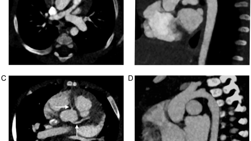
- Cardiology: Detailed imaging of small blood vessels and vascular lesions
- Neurology: Improved visualization of fine structures
- General diagnostics: Superior image quality with lower radiation doses
The Path Forward
The current clinical trials aim to:
- Evaluate deep silicon detector performance against standard CT technology
- Optimize image processing protocols
- Develop AI-driven pattern recognition capabilities
- Enhance data management and visual information systems
Clara Hellner, Chairman of MedTechLabs, expressed enthusiasm about the technology’s potential: “We’ve established a dedicated CT laboratory at Karolinska University Hospital for this clinical research. This represents an exciting first step toward technology that could benefit millions of patients worldwide.”
Jean-Luc Procaccini, President and CEO of GE Healthcare Molecular Imaging and Computed Tomography, boldly stated: “We are at the forefront of a revolution in healthcare.”
Rapid Development Timeline
GE’s swift progress is remarkable—from acquiring Prismatic Sensors AB in November 2020 to launching clinical trials in less than a year. Prismatic Sensors, originally spun off from KTH Royal Institute of Technology in Stockholm in 2012, brought cutting-edge expertise in deep silicon detector technology.
Stanford University radiology professor Norbert Pelc praised the achievement: “The scientists and engineers at Prismatic Sensors have proven they can create detectors that handle extremely high photon fluxes while maintaining excellent energy and spatial resolution—all at reasonable manufacturing costs. This represents the most significant improvement in CT technology we’ve seen in decades.”
Conclusion
As GE’s photon counting CT technology with deep silicon detectors enters clinical trials, the medical imaging field stands on the brink of transformation. This technology promises to enhance diagnostic capabilities across multiple specialties while reducing patient radiation exposure—a true win-win for healthcare providers and patients alike.