
TL;DR:
- Both standard and ultrahigh resolution modes in photon-counting computed tomography (PCCT) demonstrate high accuracy for coronary CT angiography (CCTA).
- Ultrahigh resolution mode offers an additional diagnostic advantage, particularly in patients with heavily calcified coronary arteries.
- These findings support the integration of ultrahigh resolution protocols into clinical practice for selected cases involving extensive coronary calcification.
Introduction
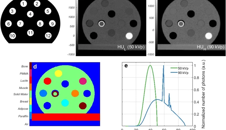
Photon-counting CT (PCCT) technology represents a significant advancement in computed tomography imaging, offering higher contrast-to-noise ratios and improved spatial resolution compared to conventional energy-integrating detector systems. This innovative technology facilitates spectral imaging and may enhance the diagnostic accuracy of cardiovascular assessments, especially in challenging cases such as extensive coronary calcification. Understanding the performance of different imaging modes within PCCT systems—particularly standard versus ultrahigh resolution—can help shape protocol optimization and improve clinical outcomes in coronary CT angiography (CCTA).
Study Overview
A recent investigation, led by Dr. Mengzhen Wang at Shanghai Jiao Tong University School of Medicine, evaluated the diagnostic performance of standard and ultrahigh resolution modes of PCCT in CCTA. The study aimed to determine whether ultrahigh resolution imaging confers an additional advantage in detecting coronary stenoses, especially in patients with severe calcification, compared to standard resolution protocols.
Methodology
The study included 122 inpatient participants who underwent CCTA between October 2023 and October 2024. Participants were divided evenly, with 61 subjects undergoing standard resolution protocols and 61 undergoing ultrahigh resolution protocols. All participants also received invasive coronary angiography (ICA), which served as the reference standard for assessing coronary stenosis.
Imaging protocols included various reconstruction techniques:
– Standard resolution: ‘SRnormal’ and ‘SRVNCa’ (virtual non-calcium) images, both reconstructed at 0.6-mm slice thickness using the Bv40 kernel.
– Ultrahigh resolution: ‘UHRnormal’ (0.6-mm slice thickness, Bv40 kernel) and ‘UHRthin’ (0.2-mm slice thickness, Bv64 kernel).
Two radiologists independently measured the diameter of identified stenoses to determine their significance, defined as a stenosis of 50% or greater.
Results and Performance Metrics
The diagnostic accuracy of the different modes was evaluated on a per-segment basis, with the following key findings:
– Standard resolution (SR normal): Sensitivity 92.9%, Specificity approximately 89–90%, Accuracy around 90%.
– Standard resolution virtual non-calcium (SRVNCa): Similar performance with sensitivity about 92.9–93.5%, specificity over 91%, and accuracy close to 92%.
– Ultrahigh resolution (UHR normal): Improved sensitivity at 96%, with specificity around 91.6–92.4%, and accuracy approximately 92–93%.
– Ultrahigh resolution thin (UHR thin): Achieved the highest performance, with sensitivity reaching 100%, specificity approximately 98.6–98.9%, and accuracy nearing 98.8–99%.
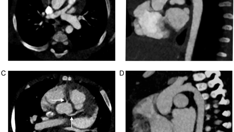
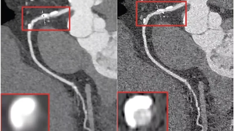
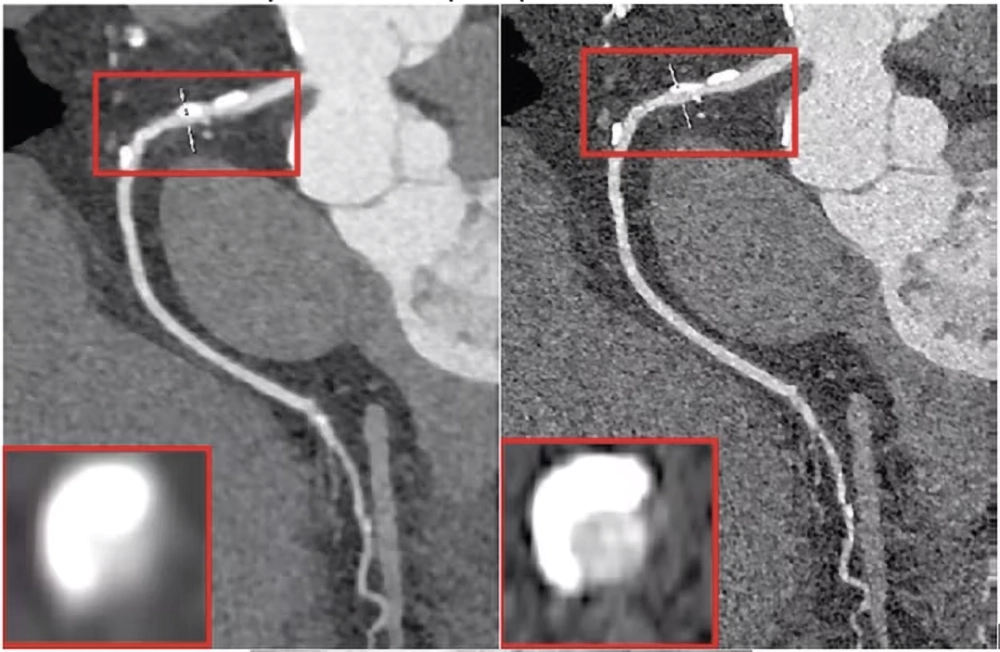
Representative cases demonstrated that ultrahigh resolution imaging reduced blooming artifacts from calcified plaques, which is a notable limitation in conventional and standard resolution CT imaging.
Significance of Findings
The study highlights that both standard resolution and ultrahigh resolution PCCT protocols achieve high diagnostic performance in detecting significant coronary stenosis, validated against invasive coronary angiography. However, the enhanced spatial resolution in ultrahigh resolution modes notably improves visualization, particularly in vessels heavily affected by calcification, reducing blooming artifacts and potentially minimizing false-positive assessments.
Implications for Clinical Practice
These findings suggest that radiology practices could consider prioritizing ultrahigh resolution modes of PCCT for patients with extensive coronary artery calcification or in cases where there is strong clinical suspicion of severe coronary artery disease (CAD). The improved spatial resolution and spectral imaging capabilities of PCCT may facilitate more precise assessment of coronary lesions and plaque characteristics, thereby informing better clinical decision-making.
Context and Technological Trends
Photon counting CT is increasingly recognized as a transformative technology in medical imaging due to its ability to acquire spectral data and enhance image quality at potentially reduced radiation doses. The integration of ultrahigh resolution modes within PCCT systems exemplifies ongoing efforts to leverage spectral imaging and improved spatial resolution for cardiovascular diagnostics. Continued research and refinement in this field are expected to expand PCCT’s clinical applications, particularly in complex cardiovascular pathologies.
Future Outlook and Conclusions
The current evidence supports the clinical utility of both standard and ultrahigh resolution PCCT modes in coronary CTA. The choice of imaging protocol should be guided by the clinical context, particularly the extent of calcification. Future studies are anticipated to further elucidate the benefits of spectral imaging, dose reduction strategies, and the development of standardized protocols for PCCT in cardiovascular imaging. As the technology matures, it has the potential to become a cornerstone in non-invasive coronary assessment, improving diagnostic accuracy and patient management.
References
The detailed findings and methodology of this study are available through the American Journal of Roentgenology and other medical imaging publications specializing in CT advancements and spectral imaging technologies.