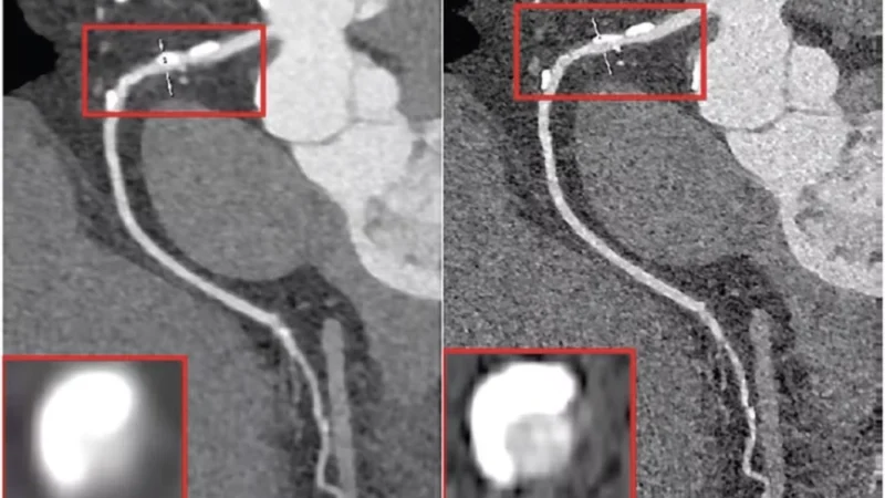

Image credits: Vaniqui, A., Schyns, L.E.J.R., Almeida, I.P. et al. The impact of dual energy CT imaging on dose calculations for pre-clinical studies. Radiat Oncol 12, 181 (2017). https://doi.org/10.1186/s13014-017-0922-9
TL;DR
- Cylindrical phantoms can overestimate accuracy in iodine quantification compared to anatomically realistic designs.
- Projection-based material decomposition is more robust and less affected by phantom geometry than image-based methods.
- Incorporating skull-mimicking materials in elliptical phantoms is essential for accurate, clinically relevant evaluation of photon-counting head CT systems.
Summary
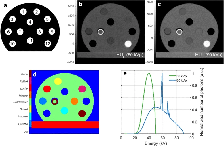
This study, published in the Journal of Medical Imaging, addresses the limitations of current evaluation practices in spectral head CT, particularly concerning iodine quantification accuracy. Traditional CT systems rely on energy-integrating detectors and dual-exposure techniques, which can increase radiation dose and introduce motion artifacts. Photon-counting detectors (PCDs) provide a promising alternative by enabling multi-energy acquisition in a single exposure with superior energy resolution. However, current evaluation standards often use cylindrical phantoms that lack anatomical realism and fail to mimic skull structures, potentially leading to overestimated system performance. To address this gap, the study compares conventional cylindrical phantoms with anatomically accurate elliptical phantoms incorporating skull-mimicking materials.
Results
Through detailed simulations using a photon-counting CT system with cadmium telluride detectors, the researchers analyzed iodine quantification accuracy under various imaging conditions. They found that image-based material decomposition methods were highly sensitive to phantom geometry, with cylindrical phantoms showing inflated accuracy and precision. In contrast, projection-based decomposition methods were more robust, maintaining consistent performance across different phantom types. The findings highlight the critical importance of using anatomically realistic phantoms in spectral CT calibration and validation to ensure clinically relevant and accurate assessments, especially for complex head imaging.