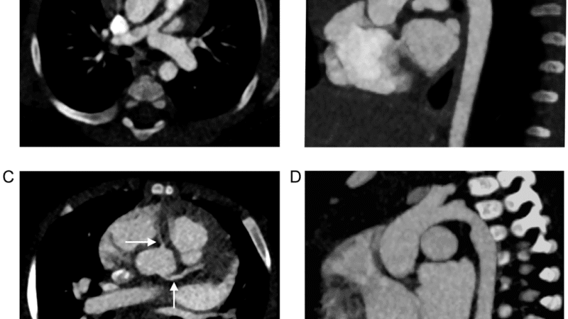

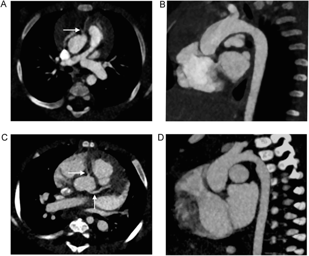
A, B Cardiac examination on photon-counting detector CT of a 7-month-old female patient, born with tetralogy of Fallot. Volume CT dose index (CTDIvol) of this examination was 0.11 mGy, dose length product (DLP) was 1.9 mGy*cm, water-equivalent diameter was 118 mm. C, D Cardiac examination on energy-integrating detector CT of a 5-month-old female patient, born with atrioventricular septal defect. CTDIvol of this examination was 0.35 mGy, DLP was 5.8 mGy*cm, water-equivalent diameter was 118 mm. The cardiac anatomy can be very well depicted in both studies, including the coronary arteries (arrows in the axial orientations A and C). B and D show the aortic arch in parasagittal orientation
TL;DR:
- Recent research demonstrates that cardiac photon-counting computed tomography (PCCT) provides similar image quality to conventional energy-integrating detector CT (EID-CT) for pediatric congenital heart disease, while reducing radiation exposure by more than 43%.
- The study reports comparable signal-to-noise and contrast-to-noise ratios for both modalities at significantly lower dose length products with PCCT.
- These findings highlight the potential of PCCT for safer imaging in pediatric cardiac patients, aligning with current trends in dose optimization and advanced spectral imaging.
Introduction
Photon-counting CT (PCCT) technology continues to reshape clinical imaging, with a growing body of evidence supporting its advantages in both image quality and radiation dose reduction. A recent study published in European Radiology evaluated the use of cardiac PCCT in pediatric patients with congenital heart disease, a population particularly sensitive to ionizing radiation. The results indicate that PCCT can maintain diagnostic image quality at substantially lower radiation doses compared to traditional energy-integrating detector CT (EID-CT).
Study Overview
The research focused on pediatric patients diagnosed with congenital heart disease who underwent cardiac CT imaging. The investigators compared the performance of PCCT and EID-CT in terms of signal-to-noise ratio (SNR), contrast-to-noise ratio (CNR), and dose length product (DLP), which is a key metric for radiation exposure. PCCT demonstrated a reduction in DLP by over 43% while yielding comparable SNR and CNR to EID-CT, indicating that lower radiation dose did not compromise image quality.
Potential Clinical Implications
Pediatric patients with congenital heart disease often require multiple imaging studies over their lifetimes, leading to cumulative radiation exposure. The demonstrated ability of PCCT to provide diagnostically equivalent imaging at significantly reduced radiation doses is an important advancement in minimizing long-term risks. This study reinforces the potential value of integrating PCCT into clinical practice, particularly for patient groups where radiation exposure is a major concern.
Takeaways
This study provides evidence that PCCT can achieve reductions in radiation dose for pediatric congenital heart disease imaging while maintaining image quality parameters. These results are aligned with ongoing trends toward dose optimization and advanced spectral imaging in clinical practice. As technology continues to evolve, photon-counting CT is poised to play an increasing role in pediatric and cardiovascular imaging.